Immunohistochemical staining of Actin, Muscle  of human FFPE tissue followed by incubation with HRP labeled secondary and development with DAB substrate.

Actin, Muscle [HHF35] Antibody (cGMP).

$ 135.00$ 495.00
Please Select Product Options Below To View The Catalog Number.

SKU: QIVD69
Species: Human
Tested Applications: IHC [IVD]
Available Conjugates:
Isotype: Mouse IgG1, kappa

Product NumberDescriptionPrice
QIVD69-0.1ml Size: 0.1 ml, Format: Concentrate $ 135.00
QIVD69-0.5ml Size: 0.5 ml, Format: Concentrate $ 295.00
QIVD69-1ml Size: 1 ml, Format: Concentrate $ 495.00
Shipping Information
In Stock
Flat Rate Shipping Anywhere in the US: $45
Datasheets and Documentation
Product Datasheet
Certificate of Analysis and Tags (Coming Soon)
Lot Number:

Expiration Date:

Concentration (Write Lyophilized if Lyophilized):

Reconsitution Instructions (Leave Blank if Liquid):

Manufacture Date:

Purity:

Bioactivity (test results eg. IU/ml):


SKU: QIVD69-0.1ml
Actin, Muscle General Information
Alternate Names
Molecular Weight
42.1 kDa
Chromosomal Location
q42.13 [chr: 1] [chr_start: 229431245] [chr_end: 229434098] [strand: -1]
Curated Database and Bioinformatic Data
Gene SymbolACTA1
Entrez Gene ID58
RefSeq Protein Accession(s)NP_001091
RefSeq mRNA Accession(s)NM_001100;
RefSeq Genomic Accession(s)NG_006672; NC_000001
UniProt ID(s)P68133
PharmGKB ID(s)PA24455
KEGG Gene ID(s)hsa:58
Associated Diseases (KEGG IDs)Nemaline myopathy 3 (NEM3) [MIM:161800]: A form of nemaline myopathy. Nemaline myopathies are muscular disorders characterized by muscle weakness of varying severity and onset, and abnormal thread-like or rod-shaped structures in muscle fibers on histologic examination. {ECO:0000269|PubMed:10508519, ECO:0000269|PubMed:11166164, ECO:0000269|PubMed:11333380, ECO:0000269|PubMed:15198992, ECO:0000269|PubMed:15236405, ECO:0000269|PubMed:15336687, ECO:0000269|PubMed:15520409, ECO:0000269|PubMed:16427282, ECO:0000269|PubMed:16945537, ECO:0000269|PubMed:17705262, ECO:0000269|PubMed:22442437, ECO:0000269|PubMed:23650303, ECO:0000269|PubMed:25938801}. The disease is caused by mutations affecting the gene represented in this entry.; Myopathy, actin, congenital, with excess of thin myofilaments (MPCETM) [MIM:161800]: A congenital muscular disorder characterized at histological level by areas of sarcoplasm devoid of normal myofibrils and mitochondria, and replaced with dense masses of thin filaments. Central cores, rods, ragged red fibers, and necrosis are absent. {ECO:0000269|PubMed:10508519}. The disease is caused by mutations affecting the gene represented in this entry.; Myopathy, congenital, with fiber-type disproportion (CFTD) [MIM:255310]: A genetically heterogeneous disorder in which there is relative hypotrophy of type 1 muscle fibers compared to type 2 fibers on skeletal muscle biopsy. However, these findings are not specific and can be found in many different myopathic and neuropathic conditions. {ECO:0000269|PubMed:15468086, ECO:0000269|PubMed:17387733}. The disease is caused by mutations affecting the gene represented in this entry.; Myopathy, scapulohumeroperoneal (SHPM) [MIM:616852]: An autosomal dominant muscular disorder characterized by progressive muscle weakness with initial scapulo-humeral-peroneal and distal distribution. Over time, muscle weakness progresses to proximal muscle groups. Clinical characteristics include scapular winging, mild lower facial weakness, foot drop due to foot eversion and dorsiflexion weakness, and selective muscle atrophy. Age at onset and disease progression are variable. {ECO:0000269|PubMed:25938801}. The disease is caused by mutations affecting the gene represented in this entry.
General Description of Actin, Muscle .
This pan actin is useful in identifying tumors arising from muscle cells, i.e. leiomyosarcoma as well as rhabdomyosarcoma.

Antibody (Suitable for clinical applications)

Sample Type: FFPE Patient Samples.
Tested Applications: IHC. Approved for In Vitro Diagnostic Procedures on FFPE tissues. For tissue collection recommendations, please see datasheet sent with product.
Application Notes
Specification Recommendation
Recommended Dilution (Conc) 1:25-1:75
Pretreatment No Pretreatment Required
Incubation Parameters 30 min at Room Temperature

Prior to use, inspect vial for the presence of any precipitate or other unusual physical properties. These can indicate that the antibody has degraded and is no longer suitable for patient samples. Please run positive and negative controls simultaneously with all patient samples to account and control for errors in laboratory procedure. Use of methods or materials not recommended by enQuire Bio including change to dilution range and detection system should be routinely validated by the user.

Clonality: Monoclonal
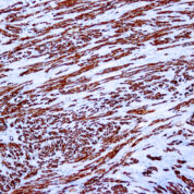
Anti-Actin, Muscle Antibody Clone: HHF35
Host and Isotype: Mouse IgG1, kappa
Recommended Positive Control Sample: Skeletal muscle
Cellular Localization of Antibody HHF35 Staining: Cytoplasmic
Buffer and Stabilizer: PBS with 1% BSA and 0.05% NaN3
Antibody Concentration: Lot specific. Plese contact tech support for data.
Immunogen: BALB/C mice were injected with SDS extracted protein from human myocardium.
Storage Conditions: This antibody should be stored refrigerated (2-8°C). This product should not be used past the expiration date printed on the vial.

Actin, Muscle Information for Pathologists

Summary:

Discovered in 1987 (Am J Pathol 1987;126:51); also called HHF35, MSA. Recognizes all alpha actins (skeletal, smooth, cardiac) and gamma smooth muscle actin; but not beta cytoplasmic or gamma cytoplasmic actin (the latter is also called non-muscle actin). Recognizes actin expressed in all cells with muscle differentiation (cardiac, smooth and skeletal muscle), myoepithelial cells, myofibroblasts, pericytes and myogenic tumors (Am J Clin Pathol 1991;96:32). Uses by pathologists Identify skeletal muscle (Tumori 2007;93:198, J Cutan Pathol 2007;34:352) and smooth muscle cells (Eur Respir J 2001;17:316) in normal tissue or various disease entities.

Common Uses By Pathologists:

Identify skeletal muscle (Tumori 2007;93:198, J Cutan Pathol 2007;34:352) and smooth muscle cells (Eur Respir J 2001;17:316) in normal tissue or various disease entities. Classify tumors with smooth or skeletal muscle, pericytes, myofibroblasts (Cardiovasc Pathol 2006;15:91) or myoepithelial cells. Differentiate leiomyosarcoma (MSA+, keratin-) from spindle cell carcinoma (MSA-, keratin+, Am J Otolaryngol 2005;26:201).

Limitations and Warranty

This antibody is manufactured in accordance with clinical good manufacturing practices in an ISO13485:2016 certified production facility. It is intended for multiple uses including in vitro diagnostic use and research use only applications. Please see vial label for expiration date. We strive to always deliver antibodies with a shelf life of at least two years.

There are no reviews yet.

Be the first to review “Actin, Muscle [HHF35] Antibody (cGMP).”

Your email address will not be published. Required fields are marked *

Share a Protocol (View Reviewed Protocols Below)

Protocol Title

Materials

Methods

Notes


  protocol submission gif
enQuire Bio, LLC   8420 S Continental Divide Rd, #202   Littleton, CO 80127
Chat: See lower right corner. | enquire@enquirebio.com | Fax: 1-720-897-3730
Customer Service Available M-F 8AM-6PM MST or 24/7 Via Email

Actin, Muscle [HHF35] Antibody (cGMP).