.jpg "Anti-Arginase 1 Specificity Assay (Array)")
.jpg)
%20Gel.jpg)
.jpg)
Human Anti-Arginase 1 Antibody Product Attributes
Species: Human
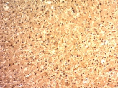
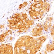
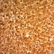
Tested Applications: Flow Cytometry, Immunofluorescence, Western Blot, Immunohistochemistry (IHC).
Application Notes: Flow Cytometry (0.5-1ug of antibody/million cells in 0.1ml), Immunofluorescence (1-2ug of antibody/ml), Western Blot (0.5-1ug of antibody/ml), Immunohistochemistry (IHC) (Formalin-fixed) (2-4ug of antibody/ml for 30 minutes at RT)
Clonality: Monoclonal
Anti-Arginase 1 Antibody Clone: ARG1/1125
Clone ARG1/1125 Host and Isotype: Mouse IgG3 kappa
Anti-Human Arginase 1 Positive Control Sample: 293T cells. Hepatocellular Carcinoma (HCC).
Cellular Localization of Antibody Cytoplasmic
Buffer and Stabilizer: 10mM PBS with 0.05% BSA & 0.05% azide.
Antibody Concentration: 200ug/ml
Antibody Purification Method:Protein A/G Purified
Immunogen: Recombinant fragment (87 Amino acid residues around aa 1-150) of human ARG1 protein
Storage Conditions: Store at 2 to 8° C (refrigerate). Stable for 24 months when properly stored.
Arginase 1 Previously Observed Antibody Staining Patterns
Observed Antibody Staining Data By Tissue Type:
Variations in Arginase 1 antibody staining intensity in immunohistochemistry on tissue sections are present across different anatomical locations. An intense signal was observed in hepatocytes in liver. More moderate antibody staining intensity was present in hepatocytes in liver. Low, but measureable presence of Arginase 1 could be seen inhematopoietic cells in the bone marrow. We were unable to detect Arginase 1 in other tissues. Disease states, inflammation, and other physiological changes can have a substantial impact on antibody staining patterns. These measurements were all taken in tissues deemed normal or from patients without known disease.Limitations and Warranty
enQuire Bio's Arginase 1 Anti-Human Monoclonal is available for Research Use Only. This antibody is guaranteed to work for a period of two years when properly stored.


-178x178.jpg)
There are no reviews yet.