

Antibody (Suitable for clinical applications)
Sample Type: FFPE Patient Samples.
Tested Applications: IHC. Approved for In Vitro Diagnostic Procedures on FFPE tissues. For tissue collection recommendations, please see datasheet sent with product.
Application Notes
| Specification | Recommendation |
|---|---|
| Recommended Dilution (Conc) | 1:50-1:100 |
| Pretreatment | Citrate Buffer pH 6.0 |
| Incubation Parameters | 30 min at Room Temperature |
Prior to use, inspect vial for the presence of any precipitate or other unusual physical properties. These can indicate that the antibody has degraded and is no longer suitable for patient samples. Please run positive and negative controls simultaneously with all patient samples to account and control for errors in laboratory procedure. Use of methods or materials not recommended by enQuire Bio including change to dilution range and detection system should be routinely validated by the user.
Clonality: Monoclonal
Anti-Calcitonin Antibody Clone: SP17
Host and Isotype: Rabbit Rabbit IgG
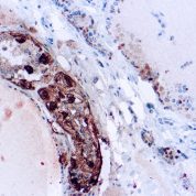
Recommended Positive Control Sample: Thyroid or medullary carcinoma
Cellular Localization of Antibody SP17 Staining: Cytoplasmic
Buffer and Stabilizer: PBS with 1% BSA and 0.05% NaN3
Antibody Concentration: Lot specific. Plese contact tech support for data.
Immunogen: Synthetic human calcitonin 1-32 amino acid peptide.
Storage Conditions: This antibody should be stored refrigerated (2-8°C). This product should not be used past the expiration date printed on the vial.
Calcitonin Information for Pathologists
Summary:
Calcitonin (thyrocalcitonin) is a 32 amino acid linear polypeptide hormone (molecular weight 3,421Kd) that participates in calcium and phosphorus metabolism. It is formed by the proteolytic cleavage of a larger pre-propeptide, which is a product of the CALC1 gene (CALC). In humans, it is primarily produced by the parafollicular cells (C cells) of the thyroid. Other tissues such as the lungs and intestinal tract can also synthesize calcitonin. The calcitonin receptor protein has been shown to be a member of the seven transmembrane G protein coupled receptor family, which is coupled by G5 to adenylate cyclase and thereby to the generation of CAMP in target cells.Common Uses By Pathologists:
As a marker for medullary thyroid carcinoma (MTC), especially when facing histologic subtypes such as the follicular, papillary or encapsulated variants that can pose diagnostic difficulties with follicular cellÂderived carcinomas and paraganglioma (Arch Pathol Lab Med 2008;132:359). Although highly specific, calcitonin staining may be patchy in medullary carcinomas and 5% of these tumors may be negative for this marker. In tumors with no staining and still suspected to be MTC, calcitonin and calcitonin geneÂrelated peptide mRNA can be demonstrated by in situ hybridization (Arch Pathol Lab Med 2010;134:207). To evaluate for C cell hyperplasia associated with familial MTC. High grade neuroendocrine tumors can also stain with calcitonin, and metastatic lesions can be misinterpreted as having arisen in the thyroid (Adv Anat Pathol 2004;11:202).Limitations and Warranty
This antibody is manufactured in accordance with clinical good manufacturing practices in an ISO13485:2016 certified production facility. It is intended for multiple uses including in vitro diagnostic use and research use only applications. Please see vial label for expiration date. We strive to always deliver antibodies with a shelf life of at least two years.


There are no reviews yet.