Human peripheral blood monocytes were stained with 5 uL APC conjugated anti-CD14 antibody (solid line) or 1 ug APC Mouse IgG1 isotype control (dashed line). Flow Cytometry Data from 10,000 events.
Human peripheral blood monocytes were stained with 5 uL APC conjugated anti-CD14 antibody (solid line) or 1 ug APC Mouse IgG1 isotype control (dashed line). Flow Cytometry Data from 10,000 events.Human peripheral blood monocytes were stained with 5 uL FITC conjugated anti-human CD14 (solid line) or 0.5 ug FITC Mouse IgG1 isotype control (dashed line). Flow Cytometry Data from 10,000 events.Human peripheral blood monocytes were stained with 5 uL  .Human peripheral blood monocytes were stained with 5 uL  (solid line) or 0.5 ug V450 Mouse IgG1 isotype control (dashed line). Flow Cytometry Data from 10,000 events.Human peripheral blood monocytes were stained with 5 uL (solid line) or 0.5 ug Qfluor™ 710 Mouse IgG1 isotype control (dashed line). Flow Cytometry Data from 10,000 events.Human peripheral blood monocytes were stained with 5 uL  (solid line) or 1 ug PE Mouse IgG1 isotype control (dashed line). Flow Cytometry Data from 10,000 events.Human peripheral blood monocytes were stained with 5 uL PerCP-Cy5.5 conjugated CD14 antibody (solid line) or 1 ug PerCP Mouse IgG1 isotype control (dashed line). Flow Cytometry Data from 10,000 events.

Anti-CD14 Antibody [61D3]

$ 88.00$ 968.00
Please Select Product Options Below To View The Catalog Number.

SKU: QAB26
Species: Human
Tested Applications: Flow Cytometry
Available Conjugates: FITC ;APC ;PerCP-Cy5.5 ;PE ;PE-Cy7 ;Qfluor 710 ;V450
Isotype: Mouse IgG1 kappa
Mass Spec Validated?: Not MS Validated

Product NumberDescriptionPrice
QAB26-F-25Tests Size: 25 tests, Tag: FITC $ 79.00
QAB26-APC-100Tests Size: 100 tests, Tag: APC $ 189.00
QAB26-APC-25Tests Size: 25 tests, Tag: APC $ 99.00
QAB26-APC-500Tests Size: 500 tests, Tag: APC $ 859.00
QAB26-F-100Tests Size: 100 tests, Tag: FITC $ 169.00
QAB26-PCP55-100Tests Size: 100 tests, Tag: PerCP-Cy5.5 $ 479.00
QAB26-PCP55-25Tests Size: 25 tests, Tag: PerCP-Cy5.5 $ 209.00
QAB26-PE-100Tests Size: 100 tests, Tag: PE $ 199.00
QAB26-PE-25Tests Size: 25 tests, Tag: PE $ 99.00
QAB26-PE7-100Tests Size: 100 tests, Tag: PE-Cy7 $ 379.00
QAB26-PE7-25Tests Size: 25 tests, Tag: PE-Cy7 $ 189.00
QAB26-QF710-100Tests Size: 100 tests, Tag: Qfluor™ 710 $ 339.00
QAB26-QF710-25Tests Size: 25 tests, Tag: Qfluor™ 710 $ 139.00
QAB26-V450-100Tests Size: 100 tests, Tag: V450 $ 229.00
QAB26-V450-25Tests Size: 25 tests, Tag: V450 $ 99.00
Shipping Information
In Stock.
Flat Rate Shipping Anywhere in the US: $45
Datasheets and Documentation
Product Datasheet
Certificate of Analysis and Tags (Coming Soon)
Lot Number:

Expiration Date:

Concentration (Write Lyophilized if Lyophilized):

Reconsitution Instructions (Leave Blank if Liquid):

Manufacture Date:

Purity:

Bioactivity (test results eg. IU/ml):


SKU: QAB26-F-25Tests
CD14 General Information
Alternate Names
mCD14
Curated Database and Bioinformatic Data
Gene SymbolCD14
Entrez Gene ID929
Ensemble Gene IDENSG00000170458
RefSeq Protein Accession(s)NP_001035110, NP_001167575, NP_001167576, NP_000582
RefSeq mRNA Accession(s)NM_001174104, NM_001040021, NM_000591, NM_001174105
RefSeq Genomic Accession(s)NG_023178, NC_018916, NC_000005
UniProt ID(s)P08571
UniGene ID(s)P08571
HGNC ID(s)1628
Cosmic ID(s)CD14
KEGG Gene ID(s)hsa:929
PharmGKB ID(s)PA26188
General Description of CD14.
Anti-human CD14 antibody, clone 61D3, is a widely used, highly published anti-CD14 clone. It binds a ~40 kDa glycoprotein identified as CD14. In the literature, the principle use of this antibody is in flow cytometry. CD14 is highly expressed on monocytes and is also present on neutrophils, several populations of macrophages, and some DCs. It may be membrane bound but is also secreted as sCD14. CD14 cooperates with TLR4 and MD2 in mediating the innate immune response to the lipopolysaccharide (LPS) which drives NFkB activation. Although best known for binding LPS, CD14 can also bind lipoteichoic acid.

Human Anti-CD14 Antibody Product Attributes

Species: Human
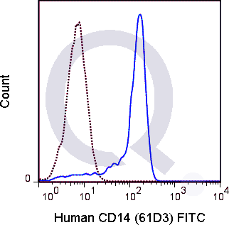
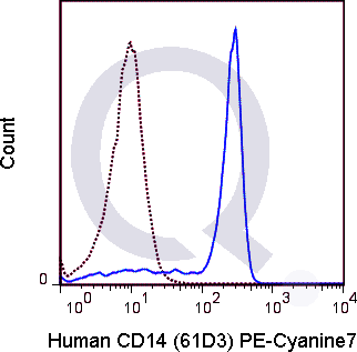
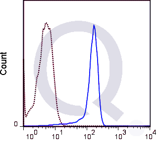
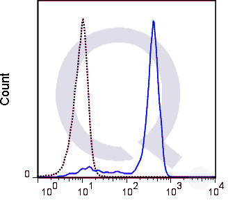
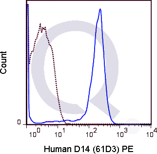
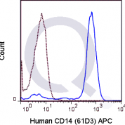
Tested Applications: Flow Cytometry.
Application Notes: 5 µl per test where one test represents staining of a cell sample in a final volume of approximately 100 µL. The number of cells within a sample must be experimentally determined, but ranges between 1x10⁵ to 1x10⁸ cells.
Clonality: Monoclonal Antibody
Anti-CD14 Antibody Clone: 61D3
Clone 61D3 Host and Isotype: Mouse IgG1 kappa
Buffer and Stabilizer: 10 mM NaH2PO4, 150 mM NaCl, 0.09% NaN3, 0.1% gelatin, pH 7.2
Antibody Concentration: 5 &microL/test
Storage Conditions: 2-8°C protected from light. Stable for 12 Months. Do Not Freeze Conjugated Formats.

CD14 Previously Observed Antibody Staining Patterns

Observed Subcellular, Organelle Specific Staining Data:

Staining with anti-CD14 antibody reveals CD14 expression is expected to be primarily localized to the plasma membrane and vesicles.

Observed Antibody Staining Data By Tissue Type:

Variations in CD14 antibody staining intensity in immunohistochemistry on tissue sections are present across different anatomical locations. An intense signal was observed in endothelial cells in the colon. More moderate antibody staining intensity was present in endothelial cells in the colon. Low, but measureable presence of CD14 could be seen in cells in the seminiferous ducts in testis, fibroblasts in skin, glandular cells in the fallopian tube and salivary gland, Langerhans in skin and Leydig cells in the testis. We were unable to detect CD14 in other tissues. Disease states, inflammation, and other physiological changes can have a substantial impact on antibody staining patterns. These measurements were all taken in tissues deemed normal or from patients without known disease.

Observed Antibody Staining Data By Tissue Disease Status:

Tissues from cancer patients, for instance, have their own distinct pattern of CD14 expression as measured by anti-CD14 antibody immunohistochemical staining. The average level of expression by tumor is summarized in the table below. The variability row represents patient to patient variability in IHC staining.

Sample Type breast cancer carcinoid cervical cancer colorectal cancer endometrial cancer glioma head and neck cancer liver cancer lung cancer lymphoma melanoma ovarian cancer pancreatic cancer prostate cancer renal cancer skin cancer stomach cancer testicular cancer thyroid cancer urothelial cancer
Signal Intensity - - - - - - - - - - - - - - - - - - - -
CD14 Variability + ++ + + + + + ++ + + + ++ ++ + + + + + + +

Selected References

Hogg N, Horton MA. 1987. In: McMichael AJ, Beverley PCL, Cobbold S, et al., eds. Leucocyte Typing III: White Cell Differentiation Antigens. New York, NY: Oxford University Press; 576-602. Haziot A, Chen S, Ferrero E, Low MG, Silber R, Goyert SM. 1988. J Immunol. 141:547-552.

Limitations and Warranty

enQuire Bio's Human Anti-CD14 Monoclonal is available for Research Use Only. This antibody is guaranteed to work for a period of two years when properly stored.

There are no reviews yet.

Be the first to review “Anti-CD14 Antibody [61D3]”

Your email address will not be published. Required fields are marked *

Share a Protocol (View Reviewed Protocols Below)

Protocol Title

Materials

Methods

Notes


enQuire Support

Flow Cytometry with anti-CD14 (61D3) of human cells from gastric cancer samples.

Materials

  • Anti-CD14 (Clone 61D3)
    • Use conjugated primary antibodies (e.g. FITC, PE, APC)
  • Mouse IgG1 Isotype Control (conjugated to the same fluorophore as 61D3).
  • Single Cell Suspension from Gastric Cancer Patient Tumor Samples
  • PBS
  • 40 μm nylon filter, 15 ml conical tubes, scissors, 3 ml syringe plunger
  • Flow cytometry staining tubes
  • Pipettes, tips, other consumables
  • FACS Canto II Flow Cytometer

Methods

  1. Prepare cell suspension from tumor samples.
    1. Using 3mm syringe plunger, mechanically disrupt tissue and press through nylon filter in the presence of PBS.
    2. Wash and resuspend cells in 1ml PBS (add 1ml of PBS, pellet cells at 400 x rcf, aspirate supernatant, and resuspend in 1ml PBS).
    3. Count cells using a hemocytometer or automated cell counter.
    4. Aliquot 1 x 10⁶ cells to separate tubes.
  2. Stain Cells with Anti-CD14 and isotype control.
    1. Wash and resuspend cells as above.
    2. Add 5 ul of Anti-CD14 to each tube containing 1×10⁶ cells. Specific dilutions here were used in an 11 color panel to distinguish tumor infiltrating lymphocytes, t-cell subsets, NK cells, and other tumor cells and should be confirmed,especially with regard to different fluorophore conjugates.
    3. Incubate samples on ice in the dark for 30 min.
    4. Wash and resuspend cells 2x as described above.
    5. Other extracellular targets may be labeled at the same time as CD14.
    6. If intracellular targets are to be stained, proceed to intracellular staining after surface marker staining is complete.
  3. Analyze Cells
    1. Cells were analyzed on a FACS Canto II Flow Cytometer.
    2. Isotype controls were run for each sample for gating and sample analysis.
    3. Compensation beads matched to the antibody (same fluorophore) were also run to allow proper measurement of each marker (this is only relevant for multicolor analyses).
    4. Additional analysis details can be viewed by visiting the publication.
6 years 3 months ago
  protocol submission gif
enQuire Bio, LLC   8420 S Continental Divide Rd, #202   Littleton, CO 80127
Chat: See lower right corner. | enquire@enquirebio.com | Fax: 1-720-897-3730
Customer Service Available M-F 8AM-6PM MST or 24/7 Via Email

Anti-CD14 Antibody [61D3]

https://enquirebio.com/wp-content/uploads/2017/11/enQuire-Bio-QAB26-APC-100Tests-anti-CD14-antibody-9-300x295.pnghttps://enquirebio.com/wp-content/uploads/2017/10/enQuire-Bio-QAB26-F-100Tests-anti-CD14-antibody-10-300x295.pnghttps://enquirebio.com/wp-content/uploads/2017/10/enQuire-Bio-QAB26-PE7-100Tests-anti-CD14-antibody-10-300x295.pnghttps://enquirebio.com/wp-content/uploads/2017/10/enQuire-Bio-QAB26-V450-100Tests-anti-CD14-antibody-10-e1515102438262-300x272.pnghttps://enquirebio.com/wp-content/uploads/2017/11/enQuire-Bio-QAB26-QF710-100Tests-anti-CD14-antibody-9-e1515102399214-300x271.pnghttps://enquirebio.com/wp-content/uploads/2017/11/enQuire-Bio-QAB26-PE-100Tests-anti-CD14-antibody-9-300x295.pnghttps://enquirebio.com/wp-content/uploads/2017/11/enQuire-Bio-QAB26-PCP55-100Tests-anti-CD14-antibody-9-300x277.png