
Mouse Anti-CD252 / OX-40L / TNFSF4 Antibody Product Attributes
Species: Mouse
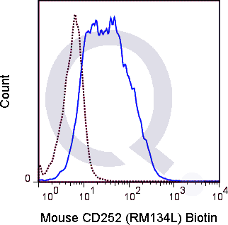
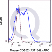
Tested Applications: Flow Cytometry.
Application Notes: See Product Datasheet for Recommended Dilution Range. Requires Experimental Optimization
Clonality: Monoclonal Antibody
Anti-CD252 / OX-40L / TNFSF4 Antibody Clone: RM134L
Clone RM134L Host and Isotype: Rat IgG2b kappa
Buffer and Stabilizer: 10 mM NaH2PO4, 150 mM NaCl, 0.09% NaN3, pH 7.2
Antibody Concentration: 0.5 mg/mL
Storage Conditions: 2-8C protected from light. Stable for 12 Months. Do Not Freeze.
CD252 / OX-40L / TNFSF4 Previously Observed Antibody Staining Patterns
Observed Subcellular, Organelle Specific Staining Data:
Staining with anti-CD252 / OX-40L / TNFSF4 antibody reveals CD252 / OX-40L / TNFSF4 expression is expected to be primarily localized to the nucleus and vesicles.Observed Antibody Staining Data By Tissue Type:
Variations in CD252 / OX-40L / TNFSF4 antibody staining intensity in immunohistochemistry on tissue sections are present across different anatomical locations. An intense signal was observed in glandular cells in the appendix, colon and duodenum, squamous epithelial cells in the esophagus, myocytes in heart muscle, squamous epithelial cells in the oral mucosa, glandular cells in the rectum and salivary gland, epidermal cells in the skin, glandular cells in the small intestine, smooth muscle cells in the smooth muscle and cells in the seminiferous ducts in testis. More moderate antibody staining intensity was present in glandular cells in the appendix, colon and duodenum, squamous epithelial cells in the esophagus, myocytes in heart muscle, squamous epithelial cells in the oral mucosa, glandular cells in the rectum and salivary gland, epidermal cells in the skin, glandular cells in the small intestine, smooth muscle cells in the smooth muscle and cells in the seminiferous ducts in testis. Low, but measureable presence of CD252 / OX-40L / TNFSF4 could be seen inlymphoid tissue in appendix, hematopoietic cells in the bone marrow, myoepithelial cells in the breast, neuronal cells in the caudate nucleus, endothelial cells in the cerebral cortex, neuronal cells in the cerebral cortex, neuropil in cerebral cortex, endothelial cells in the colon, neuronal cells in the hippocampus, cells in the glomeruli in kidney, bile duct cells in the liver, macrophages in lung, pneumocytes in lung, germinal center cells in the lymph node, follicle cells in the ovary, exocrine glandular cells in the pancreas, glandular cells in the prostate and urothelial cells in the urinary bladder. We were unable to detect CD252 / OX-40L / TNFSF4 in other tissues. Disease states, inflammation, and other physiological changes can have a substantial impact on antibody staining patterns. These measurements were all taken in tissues deemed normal or from patients without known disease.Observed Antibody Staining Data By Tissue Disease Status:
Tissues from cancer patients, for instance, have their own distinct pattern of CD252 / OX-40L / TNFSF4 expression as measured by anti-CD252 / OX-40L / TNFSF4 antibody immunohistochemical staining. The average level of expression by tumor is summarized in the table below. The variability row represents patient to patient variability in IHC staining.| Sample Type | breast cancer | carcinoid | cervical cancer | colorectal cancer | endometrial cancer | glioma | head and neck cancer | liver cancer | lung cancer | lymphoma | melanoma | ovarian cancer | pancreatic cancer | prostate cancer | renal cancer | skin cancer | stomach cancer | testicular cancer | thyroid cancer | urothelial cancer |
|---|---|---|---|---|---|---|---|---|---|---|---|---|---|---|---|---|---|---|---|---|
| Signal Intensity | - | - | - | + | + | + | - | ++ | - | - | - | - | + | ++ | - | - | + | - | + | - |
| TNFSF4 Variability | ++ | + | + | ++ | ++ | ++ | ++ | ++ | ++ | + | ++ | ++ | ++ | ++ | ++ | ++ | ++ | ++ | ++ | + |
Selected References
Akiba H, Oshima H, Takeda K, Atsuta M, Nakano H, Nakajima A, Nohara C, Yagita H and Okumura K. 1999 J Immunol. 162(12): 7058-7066. (Flow cytometry, in vitro blocking)Stuber E, Neurath M, Calderhead D, Fell HP and Strober W. 1995 Immunity. 2(5): 507-521.Sibilano R, Frossi B, Suzuki R, D'Incà F, Gri G, Piconese S, Colombo MP, Rivera J and Pucillo CE. 2012 Journal of Allergy Clin Immunol. 130(3): 751-760. (Blocking)van der Merwe M, Abdelsamed HA, Seth A, Ong T, Vogel P and Pillai AB. 2013 J Immunol. 191(11): 5764-5776 (Flow cytometry)
Limitations and Warranty
enQuire Bio's Mouse Anti-CD252 / OX40-L Monoclonal is available for Research Use Only. This antibody is guaranteed to work for a period of two years when properly stored.

There are no reviews yet.