
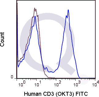
![Human PBMCs were stained with 5 uL PE conjugated anti-CD3 antibody [OKT3] (solid line) or 0.5 ug PE Mouse IgG2a isotype control (dashed line). Flow Cytometry Data from 10,000 events.](https://cdn-enquirebio.pressidium.com/wp-content/uploads/2017/10/enQuire-Bio-QAB5-PE-100Tests-anti-CD3-antibody-10.png)
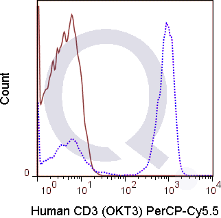
![Human PBMCs were stained with 5 uL of PE-Cy7 anti-human CD3 antibody [OKT-3] antibody and analyzed via flow cytometry.](https://cdn-enquirebio.pressidium.com/wp-content/uploads/2017/10/enQuire-Bio-QAB5-PE7-100Tests-anti-CD3-antibody-10.png)
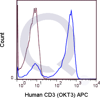
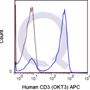
![Human PBMCs were stained with 5 uL Qfluor™ 710 conjugated anti-human CD3 antibody [clone OKT3] (solid line) or 1 ug Qfluor™ 710 Mouse IgG2a isotype control (dashed line). Flow Cytometry Data from 10,000 events.](https://cdn-enquirebio.pressidium.com/wp-content/uploads/2017/10/enQuire-Bio-QAB5-QF710-100Tests-anti-CD3-antibody-10.png)


![Human PBMCs were stained with 5 uL PE conjugated anti-CD3 antibody [OKT3] (solid line) or 0.5 ug PE Mouse IgG2a isotype control (dashed line). Flow Cytometry Data from 10,000 events.](https://cdn-enquirebio.pressidium.com/wp-content/uploads/2017/11/enQuire-Bio-QAB5-PE-100Tests-anti-CD3-antibody-9.png)
![Human PBMCs were stained with 5 uL of PE-Cy7 anti-human CD3 antibody [OKT-3] antibody and analyzed via flow cytometry.](https://cdn-enquirebio.pressidium.com/wp-content/uploads/2017/11/enQuire-Bio-QAB5-PE7-100Tests-anti-CD3-antibody-9.png)
![Human PBMCs were stained with 5 uL Qfluor™ 710 conjugated anti-human CD3 antibody [clone OKT3] (solid line) or 1 ug Qfluor™ 710 Mouse IgG2a isotype control (dashed line). Flow Cytometry Data from 10,000 events.](https://cdn-enquirebio.pressidium.com/wp-content/uploads/2017/11/enQuire-Bio-QAB5-QF710-100Tests-anti-CD3-antibody-9.png)
Human Anti-CD3 Antibody Product Attributes
Species: Human
Cross Reactivity: chimpanzee
Tested Applications: Flow Cytometry.
Application Notes: 5 ul per test where one test represents staining of a cell sample in a final volume of approximately 100 uL. The number of cells within a sample must be experimentally determined, but ranges between 1x10⁵ to 1x10⁸ cells.
Clonality: Monoclonal Antibody
Anti-CD3 Antibody Clone: OKT3
Clone OKT3 Host and Isotype: Mouse IgG2a kappa
Buffer and Stabilizer: 10 mM NaH2PO4, 150 mM NaCl, 0.09% NaN3, 0.1% gelatin, pH7.2
Antibody Concentration: 5 uL /test
Storage Conditions: 2-8C protected from light. Stable for 12 Months. Do Not Freeze.
CD3 Previously Observed Antibody Staining Patterns
Observed Antibody Staining Data By Tissue Type:
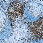
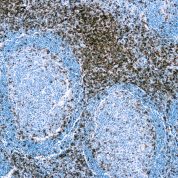
Variations in CD3 antibody staining intensity in immunohistochemistry on tissue sections are present across different anatomical locations. An intense signal was observed in lymphoid tissue in appendix and non-germinal center cells in the lymph node and tonsil. More moderate antibody staining intensity was present in lymphoid tissue in appendix and non-germinal center cells in the lymph node and tonsil. Low, but measureable presence of CD3 could be seen in. We were unable to detect CD3 in other tissues. Disease states, inflammation, and other physiological changes can have a substantial impact on antibody staining patterns. These measurements were all taken in tissues deemed normal or from patients without known disease.Selected References
Haga-Friedman A, Horovitz-Fried M, and Cohen CJ. Jun 2012. J. Immunol. 188:5538-5546. (in vitro activation)Bikker A, Moret FM, Kruize AA, Bijlsma JWJ, Lafeber FPJG, and van Roon JAG. Jun 2012. Rheumatology 51:996-1005 (in vitro activation)Bagnara D, Kaufman MS, Calissano C, et al. 2011. Blood. 117: 5463-5472. (in vivo depletion)Nguyen V, Cao L, Lin JT, Hung N, Ritz A, Yu K, Jianu R, Ulin SP, Raphael BJ, Laidlaw DH, Brossay L, and Salomon AR. 2009. Mol. Cell. Proeomics. 8: 2418-2431. (in vitro activation)Bibollet-Ruche F, McKinney BA, Duverger A, Wagner FH, Ansari AA, and Kutsch O. 2008. J. Virol. 82(20): 10271-10278. (in vitro activation - Chimpanzee)Roura-Mir C, Catalfamo M, Cheng TY, Marqusee E, Besra GS, Jaraquemada D, and Moody DB. 2005. J. Immunol. 174:3773-3780 (Immunohistochemistry - Acetone fixed frozen sections)Sato Y, Mukai K, Watanabe S, Goto M, and Shimosato Y. 1986. Am. J. Pathol. 125(3):431-435. (Immunohistochemistry - Paraffin embedded sections)
Limitations and Warranty
enQuire Bio's Human Anti-CD3 Monoclonal is available for Research Use Only. This antibody is guaranteed to work for a period of two years when properly stored.


There are no reviews yet.