
Human Anti-CD43 Antibody Product Attributes
Species: Human
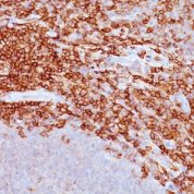
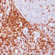
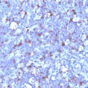
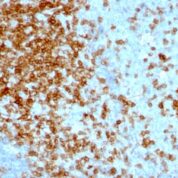
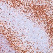
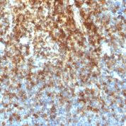
Tested Applications: Immunohistochemistry (IHC).
Clonality: Recombinant Monoclonal
Anti-CD43 Antibody Clone: SPN/1766R
Clone SPN/1766R Host and Isotype: Rabbit IgG kappa
Anti-Human CD43 Positive Control Sample: Paracortex in a tonsil or a reactive lymph node
Cellular Localization of Antibody Cell surface
Buffer and Stabilizer: 10mM PBS with or without 0.05% BSA & 0.05% azide.
Antibody Concentration: 200ug/ml
Antibody Purification Method:Protein A/G Purified
Immunogen: Recombinant full-length human SPN protein
Storage Conditions: Store at 2 to 8° C (refrigerate). Stable for 24 months when properly stored.
CD43 Previously Observed Antibody Staining Patterns
Observed Antibody Staining Data By Tissue Type:
Variations in CD43 antibody staining intensity in immunohistochemistry on tissue sections are present across different anatomical locations. An intense signal was observed in lymphoid tissue in appendix, hematopoietic cells in the bone marrow, non-germinal center cells in the lymph node, cells in the red pulp in spleen and non-germinal center cells in the tonsil. More moderate antibody staining intensity was present in lymphoid tissue in appendix, hematopoietic cells in the bone marrow, non-germinal center cells in the lymph node, cells in the red pulp in spleen and non-germinal center cells in the tonsil. Low, but measureable presence of CD43 could be seen ingerminal center cells in the lymph node, tonsil. We were unable to detect CD43 in other tissues. Disease states, inflammation, and other physiological changes can have a substantial impact on antibody staining patterns. These measurements were all taken in tissues deemed normal or from patients without known disease.Limitations and Warranty
enQuire Bio's CD43 Recombinant Monoclonal is available for Research Use Only. This antibody is guaranteed to work for a period of two years when properly stored.

There are no reviews yet.