


Human and Mouse Anti-CD44 / HCAM Antibody Product Attributes
Species: Human and Mouse
Cross Reactivity: baboon, Canine, chimpanzee, cynomolgus monkey, horse, cat, rhesus macaque, pig
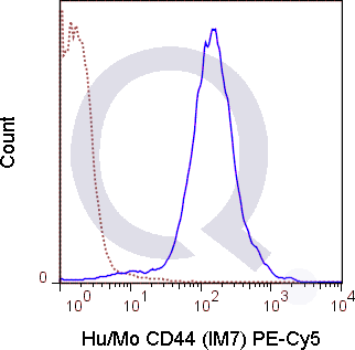
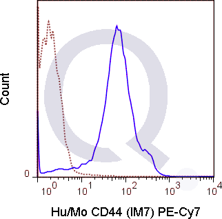
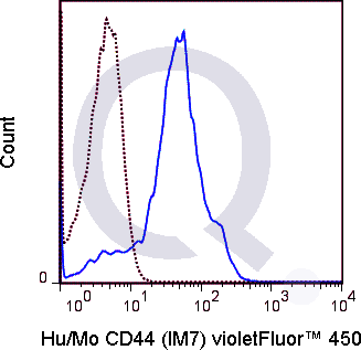
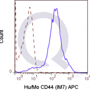
Tested Applications: Flow Cytometry.
Application Notes: See Product Datasheet for Recommended Dilution Range. Requires Experimental Optimization
Clonality: Monoclonal Antibody
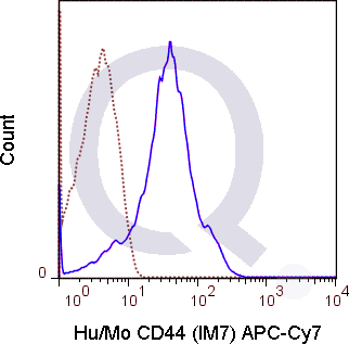
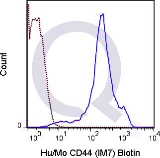
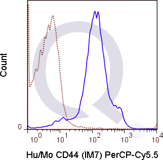
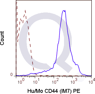
Anti-CD44 / HCAM Antibody Clone: IM7
Clone IM7 Host and Isotype: Rat IgG2b kappa
Buffer and Stabilizer: 10 mM NaH2PO4, 150 mM NaCl, 0.09% NaN3, 0.1, pH7.2
Antibody Concentration: 0.5 mg/mL
Storage Conditions: 2-8C protected from light. Stable for 12 Months. Do Not Freeze.
CD44 / HCAM Previously Observed Antibody Staining Patterns
Observed Subcellular, Organelle Specific Staining Data:
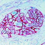
Staining with anti-CD44 / HCAM antibody reveals CD44 / HCAM expression is expected to be primarily localized to the golgi apparatus and plasma membrane.Observed Antibody Staining Data By Tissue Type:
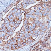
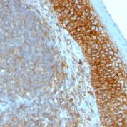
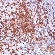
Variations in CD44 / HCAM antibody staining intensity in immunohistochemistry on tissue sections are present across different anatomical locations. An intense signal was observed in epidermal cells in the skin, glandular cells in the breast, cervix, uterine, prostate and salivary gland, hematopoietic cells in the bone marrow, keratinocytes in skin, Langerhans in skin, melanocytes in skin, respiratory epithelial cells in the bronchus and nasopharynx, squamous epithelial cells in the cervix, uterine, esophagus, oral mucosa, tonsil and vagina and urothelial cells in the urinary bladder. More moderate antibody staining intensity was present in epidermal cells in the skin, glandular cells in the breast, cervix, uterine, prostate and salivary gland, hematopoietic cells in the bone marrow, keratinocytes in skin, Langerhans in skin, melanocytes in skin, respiratory epithelial cells in the bronchus and nasopharynx, squamous epithelial cells in the cervix, uterine, esophagus, oral mucosa, tonsil and vagina and urothelial cells in the urinary bladder. Low, but measureable presence of CD44 / HCAM could be seen inadipocytes in mesenchymal tissue, cells in the glomeruli in kidney, cells in the tubules in kidney, decidual cells in the placenta, endothelial cells in the cerebral cortex and colon, follicle cells in the ovary, glandular cells in the fallopian tube, rectum and seminal vesicle, Leydig cells in the testis, neuronal cells in the cerebral cortex, neuropil in cerebral cortex, pneumocytes in lung and trophoblastic cells in the placenta. We were unable to detect CD44 / HCAM in other tissues. Disease states, inflammation, and other physiological changes can have a substantial impact on antibody staining patterns. These measurements were all taken in tissues deemed normal or from patients without known disease.Observed Antibody Staining Data By Tissue Disease Status:
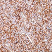
Tissues from cancer patients, for instance, have their own distinct pattern of CD44 / HCAM expression as measured by anti-CD44 / HCAM antibody immunohistochemical staining. The average level of expression by tumor is summarized in the table below. The variability row represents patient to patient variability in IHC staining.| Sample Type | breast cancer | carcinoid | cervical cancer | colorectal cancer | endometrial cancer | glioma | head and neck cancer | liver cancer | lung cancer | lymphoma | melanoma | ovarian cancer | pancreatic cancer | prostate cancer | renal cancer | skin cancer | stomach cancer | testicular cancer | thyroid cancer | urothelial cancer |
|---|---|---|---|---|---|---|---|---|---|---|---|---|---|---|---|---|---|---|---|---|
| Signal Intensity | ++ | - | +++ | ++ | ++ | +++ | +++ | + | + | +++ | +++ | + | ++ | ++ | - | ++ | - | - | ++ | - |
| CD44 Variability | ++ | + | + | ++ | ++ | ++ | + | ++ | ++ | ++ | + | ++ | ++ | ++ | ++ | ++ | ++ | ++ | ++ | ++ |
Selected References
Chandler HL, Haeussler DJ, Gemensky-Metzler AJ, Wilkie DA, and Lutz EA. 2012. Invest. Ophthalmol. Vis. Sci. 53:1835-1845. (in vitro blocking, canine)Lee L-F, Logronio K, Tu GH, Zhai W, Ni I, Mei L, Dilley J, Yu J, et al. 2012. Proc. Natl. Acad. Sci. 10.1073. (Flow cytometry).Ruffell B, Poon GFT, Lee SSM, Brown KL, Tjew S-L, Cooper J, and Johnson P. 2011. J. Biol. Chem. 286:19179-19190. (Immunoprecipitation)Miyake Y, Matsumoto H, Yokoo M, Miyazawa K et al. 2006. Biol. Reprod. 74: 501-510. (Immunohistochemistry - frozen tissue, swine)Veir JK, Lappin MR, and Dow SW. 2006. Journal of Feline Medicine and Surgery. 8:400-411. (Flow cytometry - feline)Frank NY, Margaryan A, Huang Y, Schatton T, Waaga-Gasser AM, Gasser M, Sayegh MH, Sadee W, and Frank MH. 2005. Cancer Res. 65:4320-4333. (Immunohistochemistry - frozen tissue)Fischer A, Schumacher N, Maier M, Sendtner M, and Gessler M. 2004. Genes & Dev. 18:901-911. (Immunohistochemistry - paraffin embedded tissue)Xu H, Manivannan A, Liversidge J, Sharp PF, Forrester JV, and Crane IJ. 2002. J. Leukoc. Biol. 72:1133-1141. (in vivo functional assays, induction of apoptosis)Si-Tahar M, Sitaraman S, Shibahara T, and Madara JL. 2001. Am. J. Physiol. Cell Physiol. 280:C423-C432. (in vitro functional assays, Western Blot)
Limitations and Warranty
enQuire Bio's Anti-Human/Mouse CD44 Monoclonal is available for Research Use Only. This antibody is guaranteed to work for a period of two years when properly stored.

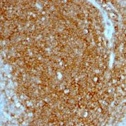
![Analysis of Mass Spec data (dashed-line) of fractions stained with CD44 / HCAM MS-QAVA™ monoclonal antibody [Clone: HCAM/918] (solid-line), reveals that less than 12.6% of signal is attributable to non-specific binding of anti-CD44 / HCAM Std. Anti-Human, Primate [Clone HCAM/918] to targets other than CD44 protein. Even frequently cited antibodies have much greater non-specific interactions, averaging over 30%. Data in image is from analysis in Jurkat, U202 and HeLa cells.](https://cdn-enquirebio.pressidium.com/wp-content/uploads/2017/10/enQuire-Bio-960-MSM2-P1-anti-CD44-HCAM-antibody-178x178.png)
There are no reviews yet.