
Human, Rat, and Zebrafish Anti-CD56 / NCAM1 Antibody Product Attributes
Species: Human, Rat, and Zebrafish
Tested Applications: Flow Cytometry, Immunofluorescence, Immunohistochemistry (IHC).
Application Notes: Flow Cytometry (0.5-1ug of antibody/million cells in 0.1ml), Immunofluorescence (0.5-1ug of antibody/ml), Immunohistochemistry (IHC) (Formalin-fixed) (0.5-1ug of antibody/ml for 30 minutes at RT)
Clonality: Monoclonal
Anti-CD56 / NCAM1 Antibody Clone: SPM128
Clone SPM128 Host and Isotype: Mouse IgG1 kappa
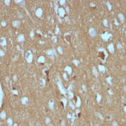
Anti-Human, Rat, and Zebrafish CD56 / NCAM1 Positive Control Sample: Cerebellum, Pancreas, Neuroblastoma
Cellular Localization of Antibody Cell surface
Buffer and Stabilizer: 10mM PBS with 0.05% BSA & 0.05% azide.
Antibody Concentration: 200ug/ml
Antibody Purification Method:Protein A/G Purified
Immunogen: Membrane preparation of a small cell lung carcinoma
Storage Conditions: Store at 2 to 8° C (refrigerate). Stable for 24 months when properly stored.
CD56 / NCAM1 Previously Observed Antibody Staining Patterns
Observed Subcellular, Organelle Specific Staining Data:
Anti-NCAM1 antibody staining is expected to be primarily localized to the cytosol and plasma membrane.Observed Antibody Staining Data By Tissue Type:
Variations in CD56 / NCAM1 antibody staining intensity in immunohistochemistry on tissue sections are present across different anatomical locations. An intense signal was observed in neuropil in cerebral cortex and peripheral nerve/ganglion in colon. More moderate antibody staining intensity was present in neuropil in cerebral cortex and peripheral nerve/ganglion in colon. Low, but measureable presence of CD56 / NCAM1 could be seen inglial cells in the hippocampus, exocrine glandular cells in the pancreas and smooth muscle cells in the smooth muscle. We were unable to detect CD56 / NCAM1 in other tissues. Disease states, inflammation, and other physiological changes can have a substantial impact on antibody staining patterns. These measurements were all taken in tissues deemed normal or from patients without known disease.Observed Antibody Staining Data By Tissue Disease Status:
Tissues from cancer patients, for instance, have their own distinct pattern of CD56 / NCAM1 expression as measured by anti-CD56 / NCAM1 antibody immunohistochemical staining. The average level of expression by tumor is summarized in the table below. The variability row represents patient to patient variability in IHC staining.| Sample Type | breast cancer | carcinoid | cervical cancer | colorectal cancer | endometrial cancer | glioma | head and neck cancer | liver cancer | lung cancer | lymphoma | melanoma | ovarian cancer | pancreatic cancer | prostate cancer | renal cancer | skin cancer | stomach cancer | testicular cancer | thyroid cancer | urothelial cancer |
|---|---|---|---|---|---|---|---|---|---|---|---|---|---|---|---|---|---|---|---|---|
| Signal Intensity | - | - | - | - | - | ++ | - | - | - | - | - | - | - | - | - | - | - | - | - | - |
| NCAM1 Variability | + | ++ | + | + | + | ++ | + | + | + | + | + | ++ | + | + | + | + | + | + | ++ | + |
Limitations and Warranty
enQuire Bio's CD56 / NCAM1 Anti-Human Monoclonal is available for Research Use Only. This antibody is guaranteed to work for a period of two years when properly stored.

There are no reviews yet.