C57Bl/6 splenocytes were stained with 0.06 ug Mouse Anti-CD62L APC (QAB49) (solid line) or 0.06 ug Rat IgG2a APC isotype control (dashed line). Flow Cytometry Data from 10,000 events.
C57Bl/6 splenocytes were stained with 0.06 ug Mouse Anti-CD62L APC (QAB49) (solid line) or 0.06 ug Rat IgG2a APC isotype control (dashed line). Flow Cytometry Data from 10,000 events.C57Bl/6 splenocytes were stained with 0.06 ug APC-Cy7 Mouse Anti-CD62L .C57Bl/6 splenocytes were stained with 0.06 ug FITC Mouse Anti-CD62L  (solid line) or 0.06 ug FITC Rat IgG2a isotype control (dashed line). Flow Cytometry Data from 10,000 events.C57Bl/6 splenocytes were stained with 0.25 ug PerCP-Cy5.5 Mouse Anti-CD62L .C57Bl/6 splenocytes were stained with 0.125 ug PE Mouse Anti-CD62L (QAB49) (solid line) or 0.125 ug PE Rat IgG2a isotype control (dashed line). Flow Cytometry Data from 10,000 events.C57Bl/6 splenocytes were stained with 0.06 ug PE-Cy7 Mouse Anti-CD62L (QAB49) (solid line) or 0.06 ug Rat IgG2a APC isotype control (dashed line). Flow Cytometry Data from 10,000 events.C57Bl/6 splenocytes were stained with 0.5 ug Qfluor™ 710 Mouse Anti-CD62L (QAB49) (solid line) or 0.5 ug  Qfluor™ 710 Rat IgG2a isotype control (dashed line). Flow Cytometry Data from 10,000 events.C57Bl/6 splenocytes were stained with 0.06 ug Mouse Anti-CD62L APC (QAB49) (solid line) or 0.06 ug Rat IgG2a APC isotype control (dashed line). Flow Cytometry Data from 10,000 events.C57Bl/6 splenocytes were stained with 0.06 ug APC-Cy7 Mouse Anti-CD62L .C57Bl/6 splenocytes were stained with 0.06 ug FITC Mouse Anti-CD62L  (solid line) or 0.06 ug FITC Rat IgG2a isotype control (dashed line). Flow Cytometry Data from 10,000 events.C57Bl/6 splenocytes were stained with 0.25 ug PerCP-Cy5.5 Mouse Anti-CD62L .C57Bl/6 splenocytes were stained with 0.125 ug PE Mouse Anti-CD62L (QAB49) (solid line) or 0.125 ug PE Rat IgG2a isotype control (dashed line). Flow Cytometry Data from 10,000 events.C57Bl/6 splenocytes were stained with 0.06 ug PE-Cy7 Mouse Anti-CD62L (QAB49) (solid line) or 0.06 ug Rat IgG2a APC isotype control (dashed line). Flow Cytometry Data from 10,000 events.C57Bl/6 splenocytes were stained with 0.5 ug Qfluor™ 710 Mouse Anti-CD62L (QAB49) (solid line) or 0.5 ug  Qfluor™ 710 Rat IgG2a isotype control (dashed line). Flow Cytometry Data from 10,000 events.

Anti-CD62L / L-Selectin Antibody [MEL-14]

$ 49.00$ 379.00
Please Select Product Options Below To View The Catalog Number.

SKU: QAB49
Species: Mouse
Tested Applications: Flow Cytometry
Available Conjugates: Biotin ;Unconjugated ;APC ;APC-Cy7 ;FITC ;PerCP-Cy5.5 ;PE ;PE-Cy7 ;Qfluor 710
Isotype: Rat IgG2a kappa
Mass Spec Validated?: Not MS Validated

Product NumberDescriptionPrice
QAB49-B-25ug Size: 25 ug, Tag: Biotin $ 49.00
QAB49-100ug Size: 100 ug, Tag: Unconjugated $ 129.00
QAB49-APC-100ug Size: 100 ug, Tag: APC $ 159.00
QAB49-APC-25ug Size: 25 ug, Tag: APC $ 59.00
QAB49-APC7-100ug Size: 100 ug, Tag: APC-Cy7 $ 309.00
QAB49-APC7-25ug Size: 25 ug, Tag: APC-Cy7 $ 109.00
QAB49-B-100ug Size: 100 ug, Tag: Biotin $ 139.00
QAB49-F-100ug Size: 100 ug, Tag: FITC $ 89.00
QAB49-F-25ug Size: 25 ug, Tag: FITC $ 59.00
QAB49-F-500ug Size: 500 ug, Tag: FITC $ 169.00
QAB49-PCP55-100ug Size: 100 ug, Tag: PerCP-Cy5.5 $ 379.00
QAB49-PCP55-25ug Size: 25 ug, Tag: PerCP-Cy5.5 $ 129.00
QAB49-PE-100ug Size: 100 ug, Tag: PE $ 169.00
QAB49-PE-25ug Size: 25 ug, Tag: PE $ 59.00
QAB49-PE7-100ug Size: 100 ug, Tag: PE-Cy7 $ 199.00
QAB49-PE7-25ug Size: 25 ug, Tag: PE-Cy7 $ 89.00
QAB49-QF710-100ug Size: 100 ug, Tag: Qfluorâ„¢ 710 $ 179.00
QAB49-QF710-25ug Size: 25 ug, Tag: Qfluorâ„¢ 710 $ 129.00
QAB49-QF630-100ug Size: 100 tests, Tag: Qfluor™ 630 $ 209.00
Shipping Information
In Stock.
Flat Rate Shipping Anywhere in the US: $45
Datasheets and Documentation
Product Datasheet
Certificate of Analysis and Tags (Coming Soon)
Lot Number:

Expiration Date:

Concentration (Write Lyophilized if Lyophilized):

Reconsitution Instructions (Leave Blank if Liquid):

Manufacture Date:

Purity:

Bioactivity (test results eg. IU/ml):


SKU: QAB49-B-25ug
CD62L General Information
Alternate Names
L-Selectin, L Selectin
Curated Database and Bioinformatic Data
Gene SymbolSell
Entrez Gene ID20343
Ensemble Gene IDENSMUSG00000026581
RefSeq Protein Accession(s)XP_006496780, NP_035476, XP_006496779, XP_006496781, NP_001157531
RefSeq mRNA Accession(s)NM_001164059, XM_006496716, XM_006496717, XM_006496718, NM_011346
RefSeq Genomic Accession(s)NC_000067
UniProt ID(s)Q3TCF3, B1B507, B1B506, P18337, Q3UV83
UniGene ID(s)Q3TCF3, B1B507, B1B506, P18337, Q3UV83
Cosmic ID(s)Sell
KEGG Gene ID(s)mmu:20343
General Description of CD62L.
The MEL-14 antibody is specific for mouse CD62L, also known as L-Selectin, a cell adhesion molecule which facilitates lymphocyte "rolling" on activated vascular endothelium and homing to high endothelial venules (HEV) as immune cells transmigrate from blood into peripheral tissues. L-Selectin is a member of a family of Selectin molecules which act together with the integrin family of adhesion molecules to mediate leukocyte-endothelial interactions. L-Selectin is characteristically expressed by neutrophils, and is also found on B cells, monocytes, granulocytes, and at varying levels on naive, effector and memory T cells. It is rapidly shed upon cell activation, releasing into the circulation a soluble form whose biological role is of particular interest in cancer biology research.The MEL-14 antibody may be used as a phenotypic marker for CD62L expression on a variety of immune cell types. Please note that CD62L (L-Selectin) itself is also referred to as MEL-14 in the literature.

Mouse Anti-CD62L Antibody Product Attributes

Species: Mouse
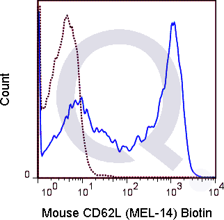
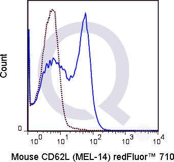
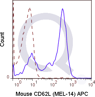
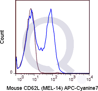
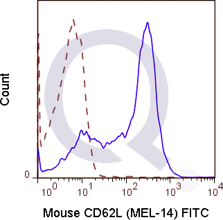
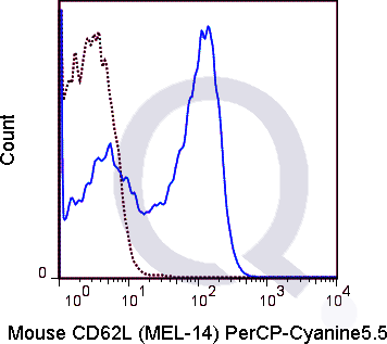
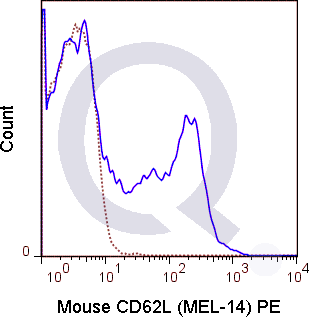
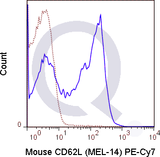
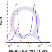
Tested Applications: Flow Cytometry.
Application Notes: See Product Datasheet for Recommended Dilution Range. Requires Experimental Optimization
Clonality: Monoclonal Antibody
Anti-CD62L Antibody Clone: MEL-14
Clone MEL-14 Host and Isotype: Rat IgG2a kappa
Buffer and Stabilizer: 10 mM NaH2PO4, 150 mM NaCl, 0.09% NaN3, pH7.2
Antibody Concentration: 0.5 mg/mL
Storage Conditions: 2-8C protected from light. Stable for 12 Months. Do Not Freeze.

CD62L Previously Observed Antibody Staining Patterns

Observed Antibody Staining Data By Tissue Type:

Variations in CD62L antibody staining intensity in immunohistochemistry on tissue sections are present across different anatomical locations. An intense signal was observed in lymphoid tissue in appendix, hematopoietic cells in the bone marrow, non-germinal center cells in the lymph node, cells in the white pulp in spleen, germinal center cells in the tonsil and non-germinal center cells in the tonsil. More moderate antibody staining intensity was present in lymphoid tissue in appendix, hematopoietic cells in the bone marrow, non-germinal center cells in the lymph node, cells in the white pulp in spleen, germinal center cells in the tonsil and non-germinal center cells in the tonsil. Low, but measureable presence of CD62L could be seen in. We were unable to detect CD62L in other tissues. Disease states, inflammation, and other physiological changes can have a substantial impact on antibody staining patterns. These measurements were all taken in tissues deemed normal or from patients without known disease.

Observed Antibody Staining Data By Tissue Disease Status:

Tissues from cancer patients, for instance, have their own distinct pattern of CD62L expression as measured by anti-CD62L antibody immunohistochemical staining. The average level of expression by tumor is summarized in the table below. The variability row represents patient to patient variability in IHC staining.

Sample Type breast cancer carcinoid cervical cancer colorectal cancer endometrial cancer glioma head and neck cancer liver cancer lung cancer lymphoma melanoma ovarian cancer pancreatic cancer prostate cancer renal cancer skin cancer stomach cancer testicular cancer thyroid cancer urothelial cancer
Signal Intensity + - + + + - + + - ++ - + + + - - - + + +
SELL Variability ++ ++ ++ ++ ++ ++ ++ + ++ ++ + + ++ + ++ ++ ++ + ++ ++

Selected References

Lee L-F, Logronio K, Tu GH, Zhai W, Ni I, Mei L, Dilley J, Yu J, et al. 2012. Proc. Natl. Acad. Sci. 10.1073. (Flow cytometry).Harp JR, Gilchrist MA, and Onami TM. 2010. J. Immunol. 185:5751-5761. (in vivo blocking)Furukawa Y, Umemoto E, Jang MH, Tohya K, Miyasaka M, and Hirata T. 2008. J. Biol. Chem. 283: 12112-12119. (Immunoelectron microscopy)Li Y, Brazzell J, Herrera A, and Walcheck B. 2006. 108: 2275-2279. (Immunoprecipitation)Ochando JC, Yopp AC, Yng Y, Garin A, Li Y, Boros P, Llodra J, Ding Y, Lira SA, Krieger NR, and Bromberg JS. 2005. J. Immunol. 174: 6993-7005. (in vivo blocking, Flow cytometry)Zhao L-C, Shey M, Farnsworth M, and Dailey MO. 2001. J. Biol. Chem. 276: 30631-30640. (Immunoprecipitation)Suzuki A, Andrew DP, Gonzalo JA, Fukumoto M, Spellberg J, Hashiyama M, Takimoto H, Gerwin N, Webb I, Molineux G, Amakawa R, Tada Y, Wakeham A, Brown J, McNiece I, Ley K, Butcher EC, Suda T, Gutierrez-Ramos JC, and Mak TK. 1996. Blood. 87:3550-3562. (Stamper-Woodruff assay; in vivo blocking)Reichert RA, Jerabek L, Gallatin WM, Butcher EC, and Weissman IL. 1986. 136(10): 3535-3542. (Immunohistochemistry)

Limitations and Warranty

enQuire Bio's Mouse Anti-CD62L / L-Selectin Monoclonal is available for Research Use Only. This antibody is guaranteed to work for a period of two years when properly stored.

There are no reviews yet.

Be the first to review “Anti-CD62L / L-Selectin Antibody [MEL-14]”

Your email address will not be published. Required fields are marked *

Share a Protocol (View Reviewed Protocols Below)

Protocol Title

Materials

Methods

Notes


  protocol submission gif
enQuire Bio, LLC   8420 S Continental Divide Rd, #202   Littleton, CO 80127
Chat: See lower right corner. | enquire@enquirebio.com | Fax: 1-720-897-3730
Customer Service Available M-F 8AM-6PM MST or 24/7 Via Email

Anti-CD62L / L-Selectin Antibody [MEL-14]

https://enquirebio.com/wp-content/uploads/2017/10/enQuire-Bio-QAB49-APC-100ug-anti-CD62L-antibody-10-292x300.pnghttps://enquirebio.com/wp-content/uploads/2017/10/enQuire-Bio-QAB49-APC7-100ug-anti-CD62L-antibody-10-300x280.pnghttps://enquirebio.com/wp-content/uploads/2017/10/enQuire-Bio-QAB49-B-100ug-anti-CD62L-antibody-10-300x295.pnghttps://enquirebio.com/wp-content/uploads/2017/10/enQuire-Bio-QAB49-F-100ug-anti-CD62L-antibody-10-300x295.pnghttps://enquirebio.com/wp-content/uploads/2017/10/enQuire-Bio-QAB49-PCP55-100ug-anti-CD62L-antibody-10-300x267.pnghttps://enquirebio.com/wp-content/uploads/2017/10/enQuire-Bio-QAB49-PE-100ug-anti-CD62L-antibody-10-292x300.pnghttps://enquirebio.com/wp-content/uploads/2017/10/enQuire-Bio-QAB49-PE7-100ug-anti-CD62L-antibody-10-300x295.pnghttps://enquirebio.com/wp-content/uploads/2017/10/enQuire-Bio-QAB49-QF710-100ug-anti-CD62L-antibody-10-300x279.pnghttps://enquirebio.com/wp-content/uploads/2017/11/enQuire-Bio-QAB49-APC-100ug-anti-CD62L-antibody-9-292x300.pnghttps://enquirebio.com/wp-content/uploads/2017/11/enQuire-Bio-QAB49-APC7-100ug-anti-CD62L-antibody-9-300x280.pnghttps://enquirebio.com/wp-content/uploads/2017/11/enQuire-Bio-QAB49-F-100ug-anti-CD62L-antibody-9-300x295.pnghttps://enquirebio.com/wp-content/uploads/2017/11/enQuire-Bio-QAB49-PCP55-100ug-anti-CD62L-antibody-9-300x267.pnghttps://enquirebio.com/wp-content/uploads/2017/11/enQuire-Bio-QAB49-PE-100ug-anti-CD62L-antibody-9-292x300.pnghttps://enquirebio.com/wp-content/uploads/2017/11/enQuire-Bio-QAB49-PE7-100ug-anti-CD62L-antibody-9-300x295.pnghttps://enquirebio.com/wp-content/uploads/2017/11/enQuire-Bio-QAB49-QF710-100ug-anti-CD62L-antibody-9-300x279.png