Immunohistochemical staining of Cytokeratin 10  of human FFPE tissue followed by incubation with HRP labeled secondary and development with DAB substrate.

Cytokeratin 10 [DE-K10] Antibody (cGMP).

$ 135.00$ 535.00
Please Select Product Options Below To View The Catalog Number.

SKU: QIVD104
Species: Human
Tested Applications: IHC [IVD]
Available Conjugates:
Isotype: Mouse IgG1, kappa

Product NumberDescriptionPrice
QIVD104-0.1ml Size: 0.1 ml, Format: Concentrate $ 135.00
QIVD104-0.5ml Size: 0.5 ml, Format: Concentrate $ 325.00
QIVD104-1ml Size: 1 ml, Format: Concentrate $ 535.00
Shipping Information
In Stock
Flat Rate Shipping Anywhere in the US: $45
Datasheets and Documentation
Product Datasheet
Certificate of Analysis and Tags (Coming Soon)
Lot Number:

Expiration Date:

Concentration (Write Lyophilized if Lyophilized):

Reconsitution Instructions (Leave Blank if Liquid):

Manufacture Date:

Purity:

Bioactivity (test results eg. IU/ml):


SKU: QIVD104-0.1ml
Cytokeratin 10 General Information
Alternate Names
Molecular Weight
58.8 kDa
Chromosomal Location
q21.2 [chr: 17] [chr_start: 40818117] [chr_end: 40822614] [strand: -1]
Curated Database and Bioinformatic Data
Gene SymbolKRT10
Entrez Gene ID3858
RefSeq Protein Accession(s)NP_000412
RefSeq mRNA Accession(s); NM_000421; XM_005257343
RefSeq Genomic Accession(s)NG_008405; NC_000017
UniProt ID(s)P13645
PharmGKB ID(s)PA30200
KEGG Gene ID(s)hsa:3858
Associated Diseases (KEGG IDs)Epidermolytic hyperkeratosis (EHK) [MIM:113800]: An autosomal dominant skin disorder characterized by widespread blistering and an ichthyotic erythroderma at birth that persist into adulthood. Histologically there is a diffuse epidermolytic degeneration in the lower spinous layer of the epidermis. Within a few weeks from birth, erythroderma and blister formation diminish and hyperkeratoses develop. {ECO:0000269|PubMed:10201536, ECO:0000269|PubMed:1380725, ECO:0000269|PubMed:1381287, ECO:0000269|PubMed:21271994, ECO:0000269|PubMed:7507150, ECO:0000269|PubMed:7507152, ECO:0000269|PubMed:7508181, ECO:0000269|PubMed:7512983, ECO:0000269|PubMed:7526210, ECO:0000269|Ref.7}. The disease is caused by mutations affecting the gene represented in this entry.; Ichthyosis annular epidermolytic (AEI) [MIM:607602]: A skin disorder resembling bullous congenital ichthyosiform erythroderma. Affected individuals present with bullous ichthyosis in early childhood and hyperkeratotic lichenified plaques in the flexural areas and extensor surfaces at later ages. The feature that distinguishes AEI from BCIE is dramatic episodes of flares of annular polycyclic plaques with scale, which coalesce to involve most of the body surface and can persist for several weeks or even months. {ECO:0000269|PubMed:9036939, ECO:0000269|PubMed:9856845}. The disease is caused by mutations affecting the gene represented in this entry.; Erythroderma, ichthyosiform, congenital reticular (CRIE) [MIM:609165]: A rare skin condition characterized by slowly enlarging islands of normal skin surrounded by erythematous ichthyotic patches in a reticulated pattern. The condition starts in infancy as a lamellar ichthyosis, with small islands of normal skin resembling confetti appearing in late childhood and at puberty. Histopathologic findings include band-like parakeratosis, psoriasiform acanthosis, and vacuolization of keratinocytes with binucleated cells in the upper epidermis, sometimes associated with amyloid deposition in the dermis. Ultrastructural abnormalities include perinuclear shells formed from a network of fine filaments in the upper epidermis. {ECO:0000269|PubMed:20798280}. The disease is caused by mutations affecting the gene represented in this entry.
General Description of Cytokeratin 10 .
This antibody recognizes keratin polypeptide of 56.5 kDa. It stains suprabasal cells in the epidermis, thymic Hassall’s bodies. Simple and glandular epithelia, transitional epithelium and adenocarcinomas are negative with this antibody.

Antibody (Suitable for clinical applications)

Sample Type: FFPE Patient Samples.
Tested Applications: IHC. Approved for In Vitro Diagnostic Procedures on FFPE tissues. For tissue collection recommendations, please see datasheet sent with product.
Application Notes
Specification Recommendation
Recommended Dilution (Conc) 1:25-1:50
Pretreatment Citrate Buffer pH 6.0
Incubation Parameters 30 min at Room Temperature

Prior to use, inspect vial for the presence of any precipitate or other unusual physical properties. These can indicate that the antibody has degraded and is no longer suitable for patient samples. Please run positive and negative controls simultaneously with all patient samples to account and control for errors in laboratory procedure. Use of methods or materials not recommended by enQuire Bio including change to dilution range and detection system should be routinely validated by the user.

Clonality: Monoclonal
Anti-Cytokeratin 10 Antibody Clone: DE-K10
Host and Isotype: Mouse IgG1, kappa
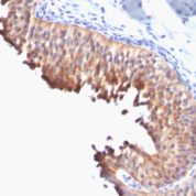
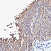
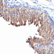
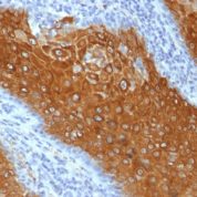
Recommended Positive Control Sample: Skin
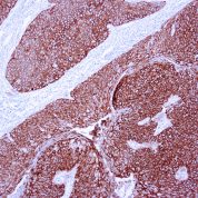
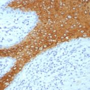
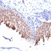
Cellular Localization of Antibody DE-K10 Staining: Cytoplasmic
Buffer and Stabilizer: PBS with 1% BSA and 0.05% NaN3
Antibody Concentration: Lot specific. Plese contact tech support for data.
Immunogen: BALB/C mice were immunized with detergent insoluble extract of human epidermis.
Storage Conditions: This antibody should be stored refrigerated (2-8°C). This product should not be used past the expiration date printed on the vial.

Cytokeratin 10 Information for Pathologists

Summary:

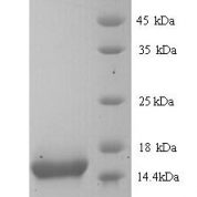
Molecular weight of 56.5 kDa. Partner of CK1. CK1 and CK10 are present in suprabasal terminally differentiating cells. Mutations in CK10 or CK1 cause epidermolytic hyperkeratosis / bullous congenital ichthyosiform erythroderma of Brocq (Hum Mol Genet 2006;15:1133, Dermatol Online J 2006;12:6); defects of CK10-CK1 protein network cause structural instability and weakness of keratinocytes, causing blistering, hyperproliferation and hyperkeratosis. CK10 is putative autoantigen in chronic, antibiotic resistant Lyme arthritis (J Immunol 2006;177:2486).

Common Uses By Pathologists:

Helps distinguish inflammatory linear verrucous epidermal nevus (higher CK10) from psoriasis (Eur J Dermatol 2004;14:216). Increase indicates a response to treatment for psoriasis (J Am Acad Dermatol 2004;51:257). Positive staining - normal Epidermal spinous and granular cell layers. Positive staining - disease

Limitations and Warranty

This antibody is manufactured in accordance with clinical good manufacturing practices in an ISO13485:2016 certified production facility. It is intended for multiple uses including in vitro diagnostic use and research use only applications. Please see vial label for expiration date. We strive to always deliver antibodies with a shelf life of at least two years.

There are no reviews yet.

Be the first to review “Cytokeratin 10 [DE-K10] Antibody (cGMP).”

Your email address will not be published. Required fields are marked *

Share a Protocol (View Reviewed Protocols Below)

Protocol Title

Materials

Methods

Notes


  protocol submission gif
enQuire Bio, LLC   8420 S Continental Divide Rd, #202   Littleton, CO 80127
Chat: See lower right corner. | enquire@enquirebio.com | Fax: 1-720-897-3730
Customer Service Available M-F 8AM-6PM MST or 24/7 Via Email

Cytokeratin 10 [DE-K10] Antibody (cGMP).