Immunohistochemical staining of Cytokeratin 14  of human FFPE tissue followed by incubation with HRP labeled secondary and development with DAB substrate.

Cytokeratin 14 [LL002] Antibody (cGMP).

$ 135.00$ 485.00
Please Select Product Options Below To View The Catalog Number.

SKU: QIVD107
Species: Human
Tested Applications: IHC [IVD]
Available Conjugates:
Isotype: Mouse IgG3

Product NumberDescriptionPrice
QIVD107-0.1ml Size: 0.1 ml, Format: Concentrate $ 135.00
QIVD107-0.5ml Size: 0.5 ml, Format: Concentrate $ 285.00
QIVD107-1ml Size: 1 ml, Format: Concentrate $ 485.00
QIVD107-6ml Size: 6 ml, Format: Predilute $ 265.00
Shipping Information
In Stock
Flat Rate Shipping Anywhere in the US: $45
Datasheets and Documentation
Product Datasheet
Certificate of Analysis and Tags (Coming Soon)
Lot Number:

Expiration Date:

Concentration (Write Lyophilized if Lyophilized):

Reconsitution Instructions (Leave Blank if Liquid):

Manufacture Date:

Purity:

Bioactivity (test results eg. IU/ml):


SKU: QIVD107-0.1ml
Cytokeratin 14 General Information
Alternate Names
Molecular Weight
51.6 kDa
Chromosomal Location
q21.2 [chr: 17] [chr_start: 41582279] [chr_end: 41586895] [strand: -1]
Curated Database and Bioinformatic Data
Gene SymbolKRT14
Entrez Gene ID3861
RefSeq Protein Accession(s)NP_000517
RefSeq mRNA Accession(s); NM_000526
RefSeq Genomic Accession(s)NG_008624; NC_000017
UniProt ID(s)P02533
PharmGKB ID(s)PA30203
KEGG Gene ID(s)hsa:3861
Associated Diseases (KEGG IDs)Epidermolysis bullosa simplex, Dowling-Meara type (DM-EBS) [MIM:131760]: A severe form of intraepidermal epidermolysis bullosa characterized by generalized herpetiform blistering, milia formation, dystrophic nails, and mucous membrane involvement. {ECO:0000269|PubMed:10583131, ECO:0000269|PubMed:10730767, ECO:0000269|PubMed:10733662, ECO:0000269|PubMed:10820403, ECO:0000269|PubMed:11710919, ECO:0000269|PubMed:12603865, ECO:0000269|PubMed:12655565, ECO:0000269|PubMed:12707098, ECO:0000269|PubMed:14987259, ECO:0000269|PubMed:16786515, ECO:0000269|PubMed:16882168, ECO:0000269|PubMed:1717157, ECO:0000269|PubMed:7561171, ECO:0000269|PubMed:7688405, ECO:0000269|PubMed:8601736, ECO:0000269|PubMed:9804355, ECO:0000269|PubMed:9989794, ECO:0000269|Ref.32}. The disease is caused by mutations affecting the gene represented in this entry.; Epidermolysis bullosa simplex, Weber-Cockayne type (WC-EBS) [MIM:131800]: A form of intraepidermal epidermolysis bullosa characterized by blistering limited to palmar and plantar areas of the skin. {ECO:0000269|PubMed:10733662, ECO:0000269|PubMed:12603865, ECO:0000269|PubMed:12655565, ECO:0000269|PubMed:12707098, ECO:0000269|PubMed:14987259, ECO:0000269|PubMed:16786515, ECO:0000269|PubMed:16882168, ECO:0000269|PubMed:7506097, ECO:0000269|PubMed:7506606, ECO:0000269|PubMed:7561171, ECO:0000269|PubMed:9284105, ECO:0000269|PubMed:9804357, ECO:0000269|PubMed:9989794}. The disease is caused by mutations affecting the gene represented in this entry.; Epidermolysis bullosa simplex, Koebner type (K-EBS) [MIM:131900]: A form of intraepidermal epidermolysis bullosa characterized by generalized skin blistering. The phenotype is not fundamentally distinct from the Dowling-Meara type, although it is less severe. {ECO:0000269|PubMed:10733662, ECO:0000269|PubMed:10820403, ECO:0000269|PubMed:11710919, ECO:0000269|PubMed:16786515, ECO:0000269|PubMed:1720261, ECO:0000269|PubMed:7526926, ECO:0000269|PubMed:7682883, ECO:0000269|PubMed:9989794, ECO:0000269|Ref.10, ECO:0000269|Ref.32}. The disease is caused by mutations affecting the gene represented in this entry.; Epidermolysis bullosa simplex, autosomal recessive 1 (EBSB1) [MIM:601001]: A form of epidermolysis bullosa, a genodermatosis characterized by recurrent blistering and cleavage within basal keratinocytes, fragility of the skin and mucosal epithelia, and erosions caused by minor mechanical trauma. {ECO:0000269|PubMed:7526933}. The disease is caused by mutations affecting the gene represented in this entry.; Naegeli-Franceschetti-Jadassohn syndrome (NFJS) [MIM:161000]: A rare autosomal dominant form of ectodermal dysplasia. The cardinal features are absence of dermatoglyphics (fingerprints), reticular cutaneous hyperpigmentation (starting at about the age of 2 years without a preceding inflammatory stage), palmoplantar keratoderma, hypohidrosis with diminished sweat gland function and discomfort provoked by heat, nail dystrophy, and tooth enamel defects. {ECO:0000269|PubMed:16960809}. The disease is caused by mutations affecting the gene represented in this entry.; Dermatopathia pigmentosa reticularis (DPR) [MIM:125595]: A rare ectodermal dysplasia characterized by lifelong persistent reticulate hyperpigmentation, non-cicatricial alopecia, and nail dystrophy. Variable features include adermatoglyphia, hypohidrosis or hyperhidrosis, and palmoplantar hyperkeratosis. {ECO:0000269|PubMed:16960809}. The disease is caused by mutations affecting the gene represented in this entry.
General Description of Cytokeratin 14 .
This antibody is specific to 50 kDa cytokeratin protein designated cytokeratin 14. Cytokeratin 14 belongs to the type A (acidic) subfamily of low molecular weight cytokeratins and exists in combination with cytokeratin 15. This antibody can be used to distinguish stratified epithelial cells from simple epithelial cells.

Antibody (Suitable for clinical applications)

Sample Type: FFPE Patient Samples.
Tested Applications: IHC. Approved for In Vitro Diagnostic Procedures on FFPE tissues. For tissue collection recommendations, please see datasheet sent with product.
Application Notes
Specification Recommendation
Recommended Dilution (Conc) 1:50-1:200
Pretreatment Citrate Buffer pH 6.0
Incubation Parameters 30 min at Room Temperature

Prior to use, inspect vial for the presence of any precipitate or other unusual physical properties. These can indicate that the antibody has degraded and is no longer suitable for patient samples. Please run positive and negative controls simultaneously with all patient samples to account and control for errors in laboratory procedure. Use of methods or materials not recommended by enQuire Bio including change to dilution range and detection system should be routinely validated by the user.

Clonality: Monoclonal
Anti-Cytokeratin 14 Antibody Clone: LL002
Host and Isotype: Mouse IgG3
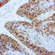
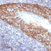
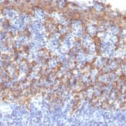
Recommended Positive Control Sample: Skin, Squamous Cell Carcinoma
Cellular Localization of Antibody LL002 Staining: Cytoplasmic
Buffer and Stabilizer: PBS with 1% BSA and 0.05% NaN3
Antibody Concentration: Lot specific. Plese contact tech support for data.
Immunogen: A synthetic peptide of 15 aa residues from the C-terminus of human keratin 14.
Storage Conditions: This antibody should be stored refrigerated (2-8°C). This product should not be used past the expiration date printed on the vial.

Cytokeratin 14 Information for Pathologists

Summary:

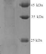
Molecular weight of 50 kDa. Partner is CK5. May be detected by cytokeratin 34BE12. CK5/6+ or CK14+ tumors define a basal subtype of DCIS (Mod Pathol 2006;19:1506) or invasive breast carcinoma; represents 9% of sporadic invasive ductal breast cancers, ER-, PR-, HER2-, high grade, poor prognosis (Mod Pathol 2005;18:1321, Eur J Cancer 2006;42:3149 but see Clin Cancer Res 2004;10:5988-not poor prognosis), associated with BRCA1 (Clin Cancer Res 2005;11:5175). In cervix, loss of expression is associated with high grade SIL and high risk HPV (Hum Pathol 2001;32:1351).

Common Uses By Pathologists:

Distinguish parathyroid oxyphil adenoma (CK14+) from carcinoma (CK14-, Am J Surg Pathol 2002;26:344). Distinguish breast papilloma (stronger and more diffuse CK14 staining) from papillary DCIS (Am J Surg Pathol 2005;29:625). Distinguish sinonasal squamous cell carcinoma (poorly differentiated or nonkeratinizing, both CK14+) from sinonasal undifferentiated carcinoma or nasopharyngeal carcinoma (CK14-, Am J Surg Pathol 2002;26:1597). Positive staining - normal Basal keratinocytes in stratified epithelium (various tissue/organs).

Limitations and Warranty

This antibody is manufactured in accordance with clinical good manufacturing practices in an ISO13485:2016 certified production facility. It is intended for multiple uses including in vitro diagnostic use and research use only applications. Please see vial label for expiration date. We strive to always deliver antibodies with a shelf life of at least two years.

There are no reviews yet.

Be the first to review “Cytokeratin 14 [LL002] Antibody (cGMP).”

Your email address will not be published. Required fields are marked *

Share a Protocol (View Reviewed Protocols Below)

Protocol Title

Materials

Methods

Notes


  protocol submission gif
enQuire Bio, LLC   8420 S Continental Divide Rd, #202   Littleton, CO 80127
Chat: See lower right corner. | enquire@enquirebio.com | Fax: 1-720-897-3730
Customer Service Available M-F 8AM-6PM MST or 24/7 Via Email

Cytokeratin 14 [LL002] Antibody (cGMP).