
Human, Pig, Mouse, and Rat Anti-Fibronectin Antibody Product Attributes
Species: Human, Pig, Mouse, and Rat
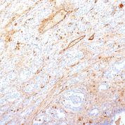
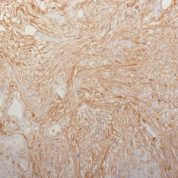
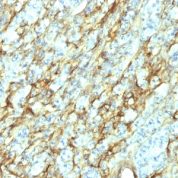
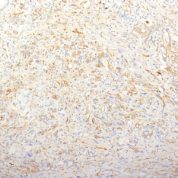
Tested Applications: Flow Cytometry, Immunofluorescence, Immunohistochemistry (IHC).
Application Notes: Flow Cytometry (0.5-1ug of antibody/million cells in 0.1ml), Immunofluorescence (0.5-1ug of antibody/ml), Immunohistochemistry (IHC) (Formalin-fixed) (0.5-1ug of antibody/ml for 30 minutes at RT)
Clonality: Monoclonal
Anti-Fibronectin Antibody Clone: 2755-8.
Clone 2755-8. Host and Isotype: Mouse IgG1 kappa
Anti-Human, Pig, Mouse, and Rat Fibronectin Positive Control Sample: SW156 cells or Kidney
Cellular Localization of Antibody <2755-8. Staining: Connective tissue matrix
Buffer and Stabilizer: 10mM PBS with 0.05% BSA & 0.05% azide.
Antibody Concentration: 200ug/ml
Antibody Purification Method:Protein A/G Purified
Immunogen: T-cell lymphoma biopsy
Storage Conditions: Store at 2 to 8° C (refrigerate). Stable for 24 months when properly stored.
Fibronectin Previously Observed Antibody Staining Patterns
Observed Antibody Staining Data By Tissue Type:
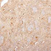
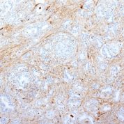
Variations in Fibronectin antibody staining intensity in immunohistochemistry on tissue sections are present across different anatomical locations. An intense signal was observed in cells in the tubules in kidney. More moderate antibody staining intensity was present in cells in the tubules in kidney. Low, but measureable presence of Fibronectin could be seen inadipocytes in breast, cells in the endometrial stroma in endometrium, cells in the molecular layer in cerebellum, cells in the red pulp in spleen, cells in the seminiferous ducts in testis, cells in the white pulp in spleen, glandular cells in the appendix, breast, epididymis, rectum, salivary gland, seminal vesicle, small intestine, stomach and thyroid gland, glial cells in the caudate nucleus, hematopoietic cells in the bone marrow, hepatocytes in liver, macrophages in lung, neuronal cells in the hippocampus, peripheral nerve in mesenchymal tissue, respiratory epithelial cells in the bronchus and nasopharynx, squamous epithelial cells in the esophagus and oral mucosa, trophoblastic cells in the placenta and urothelial cells in the urinary bladder. We were unable to detect Fibronectin in other tissues. Disease states, inflammation, and other physiological changes can have a substantial impact on antibody staining patterns. These measurements were all taken in tissues deemed normal or from patients without known disease.Observed Antibody Staining Data By Tissue Disease Status:
Tissues from cancer patients, for instance, have their own distinct pattern of Fibronectin expression as measured by anti-Fibronectin antibody immunohistochemical staining. The average level of expression by tumor is summarized in the table below. The variability row represents patient to patient variability in IHC staining.| Sample Type | breast cancer | carcinoid | cervical cancer | colorectal cancer | endometrial cancer | glioma | head and neck cancer | liver cancer | lung cancer | lymphoma | melanoma | ovarian cancer | pancreatic cancer | prostate cancer | renal cancer | skin cancer | stomach cancer | testicular cancer | thyroid cancer | urothelial cancer |
|---|---|---|---|---|---|---|---|---|---|---|---|---|---|---|---|---|---|---|---|---|
| Signal Intensity | - | - | - | + | - | - | - | ++ | + | - | - | - | ++ | - | - | - | - | + | ++ | + |
| FN1 Variability | ++ | ++ | ++ | +++ | ++ | + | ++ | +++ | ++ | + | ++ | ++ | +++ | + | + | + | ++ | ++ | ++ | +++ |
Limitations and Warranty
enQuire Bio's Fibronectin Anti-Human Monoclonal is available for Research Use Only. This antibody is guaranteed to work for a period of two years when properly stored.


There are no reviews yet.