Formalin-fixed, paraffin-embedded human Tonsil stained with HLA-DRA Monoclonal Antibody (19-26.1).

Anti-HLA-DRA Antibody Clone 19-26.1; same as MB-26.1

$ 429.00
Please Select Product Options Below To View The Catalog Number.

SKU: 3122-MSM3
Species: Human
Tested Applications: Flow Cytometry, Immunofluorescence, Immunohistochemistry (IHC)
Available Conjugates: Unconjugated
Isotype: Mouse IgG2a kappa
Mass Spec Validated?: Not MS Validated

Product NumberDescriptionPrice
3122-MSM3-P1 Size: 100 ug, Tag: Unconjugated
Shipping Information
In Stock.
Flat Rate Shipping Anywhere in the US: $45
Datasheets and Documentation
Product Datasheet
Certificate of Analysis and Tags (Coming Soon)
Lot Number:

Expiration Date:

Concentration (Write Lyophilized if Lyophilized):

Reconsitution Instructions (Leave Blank if Liquid):

Manufacture Date:

Purity:

Bioactivity (test results eg. IU/ml):


SKU: 3122-MSM3-P1
HLA-DRA General Information
Alternate Names
HLA class II histocompatibility antigen DR alpha chain, HLA-DRA
Molecular Weight
36kDa (? chain), 27kDa (? chain)
Chromosomal Location
6p21.3
Curated Database and Bioinformatic Data
Gene SymbolHLA-DRA
Entrez Gene ID3122
Ensemble Gene IDENSG00000227993, ENSG00000204287, ENSG00000277263, ENSG00000230726, ENSG00000234794, ENSG00000228987, ENSG00000226260, ENSG00000206308
RefSeq Protein Accession(s)NP_061984
RefSeq mRNA Accession(s)NM_019111
RefSeq Genomic Accession(s)NC_000006, NG_187692, NG_167247, NG_113891, NC_018917, NG_167245, NG_167246, NG_167249, NG_002392, NG_167248
UniProt ID(s)P01903, A0A0G2JMH6
UniGene ID(s)P01903, A0A0G2JMH6
HGNC ID(s)4947
Cosmic ID(s)HLA-DRA
KEGG Gene ID(s)hsa:3122
PharmGKB ID(s)PA35071
General Description of HLA-DRA.
This MAb reacts with the HLA-DR antigen, a member of MHC class II molecules. It does not cross react with HLA-DP, HLA-DQ. HLA-DR is a heterodimeric cell surface glycoprotein comprised of a 36kDa alpha (heavy) chain, a 28kDa beta (light) chain. It is expressed on B-cells, activated T-cells, monocytes/macrophages, dendritic cells, other non-professional APCs. In conjunction with the CD3/TCR complex, CD4 molecules, HLA-DR is critical for efficient peptide presentation to CD4+ T cells. It is an excellent histiocytic marker in paraffin sections producing intense cytoplasmic staining. True histiocytic neoplasms are similarly positive. HLA-DR antigens also occur on a variety of epithelial cells, their corresponding neoplastic counterparts.

Human Anti-HLA-DRA Antibody Product Attributes

Species: Human
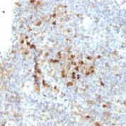
Tested Applications: Flow Cytometry, Immunofluorescence, Immunohistochemistry (IHC).
Application Notes: Flow Cytometry (0.5-1ug of antibody/million cells in 0.1ml), Immunofluorescence (0.5-1ug of antibody/ml), Immunohistochemistry (IHC) (Formalin-fixed) (1-2ug of antibody/ml for 30 minutes at RT)
Clonality: Monoclonal
Anti-HLA-DRA Antibody Clone: 19-26.1; same as MB-26.1
Clone 19-26.1; same as MB-26.1 Host and Isotype: Mouse IgG2a kappa
Anti-Human HLA-DRA Positive Control Sample: Ramos, Daudi or HuT78 cells. Tonsil or lymph node
Cellular Localization of Antibody <19-26.1; same as MB-26.1 Staining: Cell surface
Buffer and Stabilizer: 10mM PBS with 0.05% BSA & 0.05% azide.
Antibody Concentration: 200ug/ml
Antibody Purification Method:Protein A/G Purified
Immunogen: Raji Cells
Storage Conditions: Store at 2 to 8° C (refrigerate). Stable for 24 months when properly stored.

HLA-DRA Previously Observed Antibody Staining Patterns

Observed Antibody Staining Data By Tissue Type:

Variations in HLA-DRA antibody staining intensity in immunohistochemistry on tissue sections are present across different anatomical locations. An intense signal was observed in cells in the glomeruli in kidney, germinal center cells in the lymph node and tonsil, glandular cells in the duodenum and endometrium, Langerhans in skin, lymphoid tissue in appendix, macrophages in lung, myoepithelial cells in the breast and non-germinal center cells in the lymph node and tonsil. More moderate antibody staining intensity was present in cells in the glomeruli in kidney, germinal center cells in the lymph node and tonsil, glandular cells in the duodenum and endometrium, Langerhans in skin, lymphoid tissue in appendix, macrophages in lung, myoepithelial cells in the breast and non-germinal center cells in the lymph node and tonsil. Low, but measureable presence of HLA-DRA could be seen in cells in the red pulp in spleen, cells in the tubules in kidney, endothelial cells in the colon, epidermal cells in the skin, glandular cells in the appendix, cervix, uterine, epididymis and fallopian tube, glial cells in the caudate nucleus and hippocampus, myocytes in skeletal muscle, peripheral nerve in mesenchymal tissue, squamous epithelial cells in the tonsil and vagina and urothelial cells in the urinary bladder. We were unable to detect HLA-DRA in other tissues. Disease states, inflammation, and other physiological changes can have a substantial impact on antibody staining patterns. These measurements were all taken in tissues deemed normal or from patients without known disease.

Limitations and Warranty

enQuire Bio's HLA-DRA Anti-Human Monoclonal is available for Research Use Only. This antibody is guaranteed to work for a period of two years when properly stored.

There are no reviews yet.

Be the first to review “Anti-HLA-DRA Antibody Clone 19-26.1; same as MB-26.1”

Your email address will not be published. Required fields are marked *

Share a Protocol (View Reviewed Protocols Below)

Protocol Title

Materials

Methods

Notes


  protocol submission gif
enQuire Bio, LLC   8420 S Continental Divide Rd, #202   Littleton, CO 80127
Chat: See lower right corner. | enquire@enquirebio.com | Fax: 1-720-897-3730
Customer Service Available M-F 8AM-6PM MST or 24/7 Via Email

Anti-HLA-DRA Antibody Clone 19-26.1; same as MB-26.1

https://enquirebio.com/wp-content/uploads/2017/10/enQuire-Bio-3122-MSM3-P1-anti-HLA-DRA-antibody-300x225.jpeg