
Human Anti-MUC1 / EMA / CD227 Antibody Product Attributes
Species: Human
Tested Applications: Flow Cytometry, Immunofluorescence, Immunohistochemistry (IHC).
Application Notes: Flow Cytometry (0.5-1ug of antibody/million cells in 0.1ml), Immunofluorescence (1-2ug of antibody/ml), Immunohistochemistry (IHC) (Formalin-paraffin) (0.25-0.5ug of antibody/ml for 30 minutes at RT)
Clonality: Monoclonal
Anti-MUC1 / EMA / CD227 Antibody Clone: HMPV
Clone HMPV Host and Isotype: Mouse IgG1 kappa
Anti-Human MUC1 / EMA / CD227 Positive Control Sample: MCF-7 or MDA-231 cells. Breast, colon, ovarian, endometrial carcinoma.
Cellular Localization of Antibody Cytoplasmic, cell surface
Buffer and Stabilizer: 10mM PBS with 0.05% BSA & 0.05% azide.
Antibody Concentration: 200ug/ml
Antibody Purification Method:Protein A/G Purified
Immunogen: Human breast cancer cell line ZR-75 cells
Storage Conditions: Store at 2 to 8° C (refrigerate). Stable for 24 months when properly stored.
MUC1 / EMA / CD227 Previously Observed Antibody Staining Patterns
Observed Subcellular, Organelle Specific Staining Data:
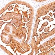
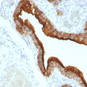
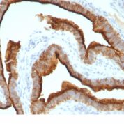
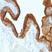
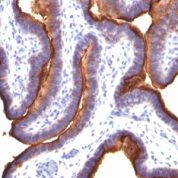
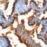
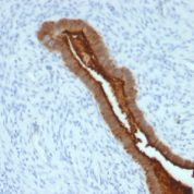
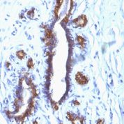
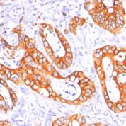
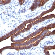
Anti-MUC1 antibody staining is expected to be primarily localized to the plasma membrane and vesicles.Observed Antibody Staining Data By Tissue Type:
Variations in MUC1 / EMA / CD227 antibody staining intensity in immunohistochemistry on tissue sections are present across different anatomical locations. An intense signal was observed in respiratory epithelial cells in the bronchus, glandular cells in the cervix, uterine, colon, endometrium, fallopian tube and gallbladder, respiratory epithelial cells in the nasopharynx and glandular cells in the rectum and stomach. More moderate antibody staining intensity was present in respiratory epithelial cells in the bronchus, glandular cells in the cervix, uterine, colon, endometrium, fallopian tube and gallbladder, respiratory epithelial cells in the nasopharynx and glandular cells in the rectum and stomach. Low, but measureable presence of MUC1 / EMA / CD227 could be seen inglandular cells in the adrenal gland, hematopoietic cells in the bone marrow, myoepithelial cells in the breast, squamous epithelial cells in the cervix, uterine, macrophages in lung, non-germinal center cells in the lymph node, squamous epithelial cells in the oral mucosa, glandular cells in the prostate and salivary gland, fibroblasts in skin, keratinocytes in skin, Langerhans in skin, melanocytes in skin, epidermal cells in the skin, cells in the seminiferous ducts in testis, Leydig cells in the testis and squamous epithelial cells in the tonsil. We were unable to detect MUC1 / EMA / CD227 in other tissues. Disease states, inflammation, and other physiological changes can have a substantial impact on antibody staining patterns. These measurements were all taken in tissues deemed normal or from patients without known disease.Observed Antibody Staining Data By Tissue Disease Status:
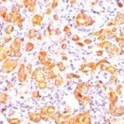
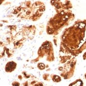
Tissues from cancer patients, for instance, have their own distinct pattern of MUC1 / EMA / CD227 expression as measured by anti-MUC1 / EMA / CD227 antibody immunohistochemical staining. The average level of expression by tumor is summarized in the table below. The variability row represents patient to patient variability in IHC staining.| Sample Type | breast cancer | carcinoid | cervical cancer | colorectal cancer | endometrial cancer | glioma | head and neck cancer | liver cancer | lung cancer | lymphoma | melanoma | ovarian cancer | pancreatic cancer | prostate cancer | renal cancer | skin cancer | stomach cancer | testicular cancer | thyroid cancer | urothelial cancer |
|---|---|---|---|---|---|---|---|---|---|---|---|---|---|---|---|---|---|---|---|---|
| Signal Intensity | +++ | - | +++ | ++ | +++ | - | +++ | - | +++ | - | - | +++ | +++ | - | +++ | - | +++ | - | ++ | ++ |
| MUC1 Variability | + | ++ | + | ++ | + | + | ++ | ++ | ++ | + | + | + | ++ | + | ++ | ++ | + | ++ | ++ | ++ |
Limitations and Warranty
enQuire Bio's MUC1 / EMA / CD227 Anti-Human Monoclonal is available for Research Use Only. This antibody is guaranteed to work for a period of two years when properly stored.

There are no reviews yet.