
Anti-Myogenin Antibody Product Attributes
Species: Human, Mouse,and Rat
Predicted Cross-Reactivity: Canine, Cat, and Pig.
Tested Applications: ELISA, Flow Cytometry, Immunofluorescence, Western Blot, Immunoprecipitation, Immunohistochemistry (IHC).
Application Notes: ELISA, Flow Cytometry (0.5-1ug of antibody/million cells in 0.1ml), Immunofluorescence (0.5-1ug of antibody/ml), Western Blot (0.5-1.0ug of antibody/ml), Immunoprecipitation (0.5-1 ug of antibody/500ug protein Lysate), Immunohistochemistry (IHC) (Formalin-fixed) (0.5-1.0ug of antibody/ml for 30 minutes at RT)
Clonality: Monoclonal
Anti-Myogenin Antibody Clone: F5D
Clone F5D Host and Isotype: Mouse IgG1 kappa
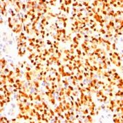
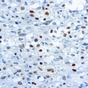
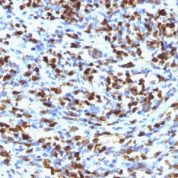
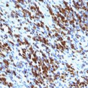
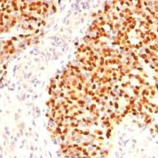
Myogenin Antibody Positive Control Samples: Rh-30 cells. Skeletal muscle or rhabdomyosarcoma.
Cellular Localization of Antibody F5D Staining: Nuclear
Buffer and Stabilizer: 10mM PBS with 0.05% BSA & 0.05% azide.
Antibody Concentration: 200ug/ml
Antibody Purification Method:Protein A/G Purified
Immunogen: Rat myogenin peptide (aa 73-94) followed by rat myogenin recombinant fragment (aa30-224) (Epitope aa138-158)
Storage Conditions: Store at 2 to 8° C (refrigerate). Stable for 24 months when properly stored.
Myogenin Previously Observed Antibody Staining Patterns
Observed Subcellular, Organelle Specific Staining Data:
Anti-MYOG antibody staining is expected to be primarily localized to the nucleoplasm. There is variability in either the signal strength or the localization of signal in nucleoplasm from cell to cell.Observed Antibody Staining Data By Tissue Disease Status:
Tissues from cancer patients, for instance, have their own distinct pattern of Myogenin expression as measured by anti-Myogenin antibody immunohistochemical staining. The average level of expression by tumor is summarized in the table below. The variability row represents patient to patient variability in IHC staining.| Sample Type | breast cancer | carcinoid | cervical cancer | colorectal cancer | endometrial cancer | glioma | head and neck cancer | liver cancer | lung cancer | lymphoma | melanoma | ovarian cancer | pancreatic cancer | prostate cancer | renal cancer | skin cancer | stomach cancer | testicular cancer | thyroid cancer | urothelial cancer |
|---|---|---|---|---|---|---|---|---|---|---|---|---|---|---|---|---|---|---|---|---|
| Signal Intensity | + | + | - | - | - | + | + | + | - | + | + | ++ | - | + | + | - | - | + | - | - |
| MYOG Variability | ++ | ++ | ++ | + | ++ | ++ | ++ | ++ | + | ++ | ++ | ++ | ++ | ++ | +++ | ++ | ++ | ++ | ++ | ++ |
Limitations and Warranty
enQuire Bio's Myogenin Anti-Human Monoclonal is available for Research Use Only. This antibody is guaranteed to work for a period of two years when properly stored.

There are no reviews yet.