
Human and Mouse Anti-p57Kip2 Antibody Product Attributes
Species: Human and Mouse
Tested Applications: Flow Cytometry, Immunofluorescence, Immunohistochemistry (IHC).
Application Notes: Flow Cytometry (0.5-1ug of antibody/million cells in 0.1ml), Immunofluorescence (0.5-1ug of antibody/ml), Immunohistochemistry (IHC) (Formalin-fixed) (0.25-0.5ug of antibody/ml for 30 minutes at RT)
Clonality: Monoclonal
Anti-p57Kip2 Antibody Clone: KIP2/880
Clone KIP2/880 Host and Isotype: Mouse IgG2b kappa
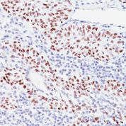
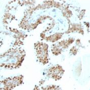
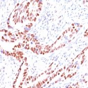
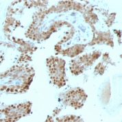
Anti-Human and Mouse p57Kip2 Positive Control Sample: LS174T, Raji, HT29, SK-BR3 cells. Colon or Prostate carcinomas.
Cellular Localization of Antibody Nuclear
Buffer and Stabilizer: 10mM PBS with 0.05% BSA & 0.05% azide.
Antibody Concentration: 200ug/ml
Antibody Purification Method:Protein A/G Purified
Immunogen: Recombinant human p57Kip2 protein
Storage Conditions: Store at 2 to 8° C (refrigerate). Stable for 24 months when properly stored.
p57Kip2 Previously Observed Antibody Staining Patterns
Observed Subcellular, Organelle Specific Staining Data:
Anti-CDKN1C antibody staining is expected to be primarily localized to the cytosol, nuclear bodies and nucleoplasm.Observed Antibody Staining Data By Tissue Type:
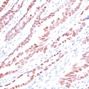
Variations in p57Kip2 antibody staining intensity in immunohistochemistry on tissue sections are present across different anatomical locations. An intense signal was observed in cells in the glomeruli in kidney, decidual cells in the placenta and trophoblastic cells in the placenta. More moderate antibody staining intensity was present in cells in the glomeruli in kidney, decidual cells in the placenta and trophoblastic cells in the placenta. Low, but measureable presence of p57Kip2 could be seen inglandular cells in the duodenum and small intestine and islets of Langerhans in pancreas. We were unable to detect p57Kip2 in other tissues. Disease states, inflammation, and other physiological changes can have a substantial impact on antibody staining patterns. These measurements were all taken in tissues deemed normal or from patients without known disease.Observed Antibody Staining Data By Tissue Disease Status:
Tissues from cancer patients, for instance, have their own distinct pattern of p57Kip2 expression as measured by anti-p57Kip2 antibody immunohistochemical staining. The average level of expression by tumor is summarized in the table below. The variability row represents patient to patient variability in IHC staining.| Sample Type | breast cancer | carcinoid | cervical cancer | colorectal cancer | endometrial cancer | glioma | head and neck cancer | liver cancer | lung cancer | lymphoma | melanoma | ovarian cancer | pancreatic cancer | prostate cancer | renal cancer | skin cancer | stomach cancer | testicular cancer | thyroid cancer | urothelial cancer |
|---|---|---|---|---|---|---|---|---|---|---|---|---|---|---|---|---|---|---|---|---|
| Signal Intensity | + | - | + | - | - | + | - | - | + | - | - | + | - | - | - | + | - | + | + | + |
| CDKN1C Variability | ++ | ++ | ++ | ++ | ++ | ++ | ++ | ++ | ++ | + | ++ | ++ | ++ | ++ | ++ | ++ | ++ | ++ | ++ | ++ |
Limitations and Warranty
enQuire Bio's p57Kip2 Anti-Human, Mouse Monoclonal is available for Research Use Only. This antibody is guaranteed to work for a period of two years when properly stored.

There are no reviews yet.