
Human, Monkey, Pig, Mouse, Rat, Chicken, Zebrafish, Drosophila melanogaster, and Yeast (S pombe & S cerevisiae) Anti-PCNA Antibody Product Attributes
Species: Human, Monkey, Pig, Mouse, Rat, Chicken, Zebrafish, Drosophila melanogaster, and Yeast (S pombe & S cerevisiae)
Tested Applications: Flow Cytometry, Immunofluorescence, Western Blot, Immunohistochemistry (IHC).
Application Notes: Flow Cytometry (0.5-1ug of antibody/million cells in 0.1ml), Immunofluorescence (0.5-1ug of antibody/ml), Western Blot (0.5-1ug of antibody/ml), Immunohistochemistry (IHC) (Formalin-fixed) (0.25-0.5ug of antibody/ml for 30 minutes at RT)
Clonality: Monoclonal
Anti-PCNA Antibody Clone: SPM350
Clone SPM350 Host and Isotype: Mouse IgG2a kappa
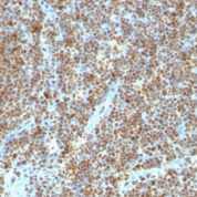
Anti-Human, Monkey, Pig, Mouse, Rat, Chicken, Zebrafish, Drosophila melanogaster, and Yeast (S pombe & S cerevisiae) PCNA Positive Control Sample: Tonsil or reactive lymph node
Cellular Localization of Antibody Predominantly nuclear, some cytoplasmic
Buffer and Stabilizer: 10mM PBS with 0.05% BSA & 0.05% azide.
Antibody Concentration: 200ug/ml
Antibody Purification Method:Protein A/G Purified
Immunogen: Rat PCNA/Protein A fusion protein
Storage Conditions: Store at 2 to 8° C (refrigerate). Stable for 24 months when properly stored.
PCNA Previously Observed Antibody Staining Patterns
Observed Subcellular, Organelle Specific Staining Data:
Anti-PCNA antibody staining is expected to be primarily localized to the nuclear bodies and nucleoplasm. There is variability in either the signal strength or the localization of signal in nuclear bodies and nucleoplasm from cell to cell.Observed Antibody Staining Data By Tissue Type:
Variations in PCNA antibody staining intensity in immunohistochemistry on tissue sections are present across different anatomical locations. An intense signal was observed in glandular cells in the appendix, lymphoid tissue in appendix, hematopoietic cells in the bone marrow, glandular cells in the colon, germinal center cells in the lymph node, glandular cells in the small intestine and germinal center cells in the tonsil. More moderate antibody staining intensity was present in glandular cells in the appendix, lymphoid tissue in appendix, hematopoietic cells in the bone marrow, glandular cells in the colon, germinal center cells in the lymph node, glandular cells in the small intestine and germinal center cells in the tonsil. Low, but measureable presence of PCNA could be seen inglandular cells in the adrenal gland, cells in the granular layer in cerebellum, cells in the molecular layer in cerebellum, myocytes in heart muscle, macrophages in lung, pneumocytes in lung, respiratory epithelial cells in the nasopharynx, exocrine glandular cells in the pancreas, glandular cells in the parathyroid gland, trophoblastic cells in the placenta, myocytes in skeletal muscle, Langerhans in skin, smooth muscle cells in the smooth muscle, cells in the red pulp in spleen and glandular cells in the stomach. We were unable to detect PCNA in other tissues. Disease states, inflammation, and other physiological changes can have a substantial impact on antibody staining patterns. These measurements were all taken in tissues deemed normal or from patients without known disease.Observed Antibody Staining Data By Tissue Disease Status:
Tissues from cancer patients, for instance, have their own distinct pattern of PCNA expression as measured by anti-PCNA antibody immunohistochemical staining. The average level of expression by tumor is summarized in the table below. The variability row represents patient to patient variability in IHC staining.| Sample Type | breast cancer | carcinoid | cervical cancer | colorectal cancer | endometrial cancer | glioma | head and neck cancer | liver cancer | lung cancer | lymphoma | melanoma | ovarian cancer | pancreatic cancer | prostate cancer | renal cancer | skin cancer | stomach cancer | testicular cancer | thyroid cancer | urothelial cancer |
|---|---|---|---|---|---|---|---|---|---|---|---|---|---|---|---|---|---|---|---|---|
| Signal Intensity | +++ | ++ | +++ | +++ | ++ | - | +++ | ++ | +++ | ++ | +++ | ++ | +++ | + | ++ | +++ | ++ | ++ | + | +++ |
| PCNA Variability | ++ | ++ | + | ++ | +++ | ++ | ++ | +++ | + | ++ | ++ | ++ | ++ | ++ | ++ | ++ | ++ | ++ | ++ | ++ |
Limitations and Warranty
enQuire Bio's PCNA Anti-Human Monoclonal is available for Research Use Only. This antibody is guaranteed to work for a period of two years when properly stored.

![Analysis of Mass Spec data (dashed-line) of fractions stained with PCNA MS-QAVA™ monoclonal antibody [Clone: PC10] (solid-line), reveals that less than 3.7% of signal is attributable to non-specific binding of anti-PCNA [Clone PC10] to targets other than PCNA protein. Even frequently cited antibodies have much greater non-specific interactions, averaging over 30%. Data in image is from analysis in A431, RT4 and MCF7 cells.](https://cdn-enquirebio.pressidium.com/wp-content/uploads/2017/10/enQuire-Bio-5111-MSM1-P1-anti-PCNA-antibody-178x178.png)

There are no reviews yet.