

Antibody (Suitable for clinical applications)
Sample Type: FFPE Patient Samples.
Tested Applications: IHC. Approved for In Vitro Diagnostic Procedures on FFPE tissues. For tissue collection recommendations, please see datasheet sent with product.
Application Notes
| Specification | Recommendation |
|---|---|
| Recommended Dilution (Conc) | 1:50-1:100 |
| Pretreatment | EDTA Buffer pH 8.0 |
| Incubation Parameters | 30 min at Room Temperature |
Prior to use, inspect vial for the presence of any precipitate or other unusual physical properties. These can indicate that the antibody has degraded and is no longer suitable for patient samples. Please run positive and negative controls simultaneously with all patient samples to account and control for errors in laboratory procedure. Use of methods or materials not recommended by enQuire Bio including change to dilution range and detection system should be routinely validated by the user.
Clonality: Polyclonal
Anti-SOX-10 Antibody Clone: Polyclonal
Host and Isotype: Rabbit Undetermined
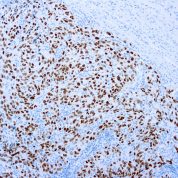
Recommended Positive Control Sample: Melanoma
Cellular Localization of Antibody Polyclonal Staining: Nuclear
Buffer and Stabilizer: PBS with 1% BSA and 0.05% NaN3
Antibody Concentration: Lot specific. Plese contact tech support for data.
Immunogen: Recombinant fragment corresponding to residues in human SOX-10 protein
Storage Conditions: This antibody should be stored refrigerated (2-8°C). This product should not be used past the expiration date printed on the vial.
SOX-10 Information for Pathologists
Summary:
SRY-related HMG-box 10 (SOX10) protein. Transcription factor known to be crucial in the specification of the neural crest and maintenance of Schwann cells and melanocytes. Expressed in nuclei of melanocytes, breast myoepithelial cells. Clinical features Usually positive nuclear staining in breast basal-like / unclassified triple negative / metaplastic carcinoma (Hum Pathol 2013;44:959).Notable Clinical Features:
Usually positive nuclear staining in breast basal-like / unclassified triple negative / metaplastic carcinoma (Hum Pathol 2013;44:959). Usually negative / rarely positive staining in breast luminal A/B or HER2+ carcinoma (5% positive), ER-/PR- DCIS (4% positive), phyllodes tumor, fibroadenoma. Specific (93%), but not very sensitive (67%) for malignant peripheral nerve sheath tumor over synovial sarcoma (Mod Pathol 2014;27:55). Must interpret with caution in intraneural tumors. In major salivary glands, expressed in nuclei of acini and both luminal and abluminal cells of intercalated ducts, but not in other sites (Mod Pathol 2013;26:1041).Common Uses By Pathologists:
Melanoma marker (100%, Appl Immunohistochem Mol Morphol 2014;22:142). Nevi are positive (Nat Cell Biol 2012;14:882), desmoplastic melanoma may be only focally positive (Appl Immunohistochem Mol Morphol 2013;21:506). SOX10 shows an increased specificity for soft tissue tumors of neural crest origin compared with S100 (Appl Immunohistochem Mol Morphol 2012;20:445). MITF1 and SOX10 can be used to differentiate melanoma in situ from actinic keratosis with melanocytic hyperplasia (Am J Dermatopathol 2014;36:124). Microscopic (histologic) imagesLimitations and Warranty
This antibody is manufactured in accordance with clinical good manufacturing practices in an ISO13485:2016 certified production facility. It is intended for multiple uses including in vitro diagnostic use and research use only applications. Please see vial label for expiration date. We strive to always deliver antibodies with a shelf life of at least two years.

There are no reviews yet.