.jpg "Anti-Aurora B Specificity Assay (Array)")
.jpg)
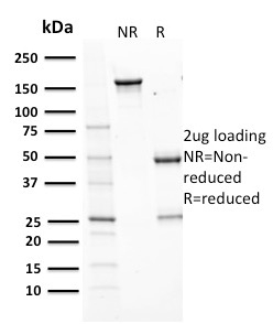
%20SDS-PAGE%20Gel.jpg)
Human Anti-Aurora B (Proliferation Marker) Antibody Product Attributes
Species: Human
Tested Applications: IHC.
Clonality: Monoclonal
Anti-Aurora B (Proliferation Marker) Antibody Clone: AURKB/1593
Clone AURKB/1593 Host and Isotype: Mouse IgG1
Anti-Human Aurora B (Proliferation Marker) Positive Control Sample: HeLa cells. Tonsil, Placenta, Hodgkin s Lymphoma or Colon Carcinoma
Cellular Localization of Antibody AURKB/1593 Staining: Nuclear and Cytoplasmic
Buffer and Stabilizer: 10mM PBS with 0.05% BSA & 0.05% azide. Also available without BSA & Azide.
Antibody Concentration: 200 ug/ml
Antibody Purification Method:Protein A/G purified from Bioreactor concentrate.
Immunogen: Recombinant human Aurora B protein fragment (aa89-251)
Storage Conditions: Store at 2 to 8 C (refrigerate). Stable for 24 months when properly stored.
Aurora B (Proliferation Marker) Previously Observed Antibody Staining Patterns
Observed Subcellular, Organelle Specific Staining Data:
Anti-AURKB antibody staining is expected to be primarily localized to the midbody and nucleoplasm. There is variability in either the signal strength or the localization of signal in midbody and nucleoplasm from cell to cell. Additionally, antibody labeling in the midbody is dependent upon phase within the cell cycle.Observed Antibody Staining Data By Tissue Type:
Variations in Aurora B antibody staining intensity in immunohistochemistry on tissue sections are present across different anatomical locations. Low, but measureable presence of Aurora B could be seen in cells in the endometrial stroma in endometrium, cells in the white pulp in spleen, decidual cells in the placenta, epidermal cells in the skin, glandular cells in the adrenal gland, breast, colon, duodenum, endometrium, gallbladder, small intestine and stomach, glial cells in the cerebral cortex, keratinocytes in skin, macrophages in lung, myocytes in skeletal muscle, neuronal cells in the cerebral cortex, respiratory epithelial cells in the bronchus, smooth muscle cells in the smooth muscle, squamous epithelial cells in the esophagus and oral mucosa, trophoblastic cells in the placenta and urothelial cells in the urinary bladder. We were unable to detect Aurora B in other tissues. Disease states, inflammation, and other physiological changes can have a substantial impact on antibody staining patterns. These measurements were all taken in tissues deemed normal or from patients without known disease.Observed Antibody Staining Data By Tissue Disease Status:
Tissues from cancer patients, for instance, have their own distinct pattern of Aurora B expression as measured by anti-Aurora B antibody immunohistochemical staining. The average level of expression by tumor is summarized in the table below. The variability row represents patient to patient variability in IHC staining.| Sample Type | breast cancer | carcinoid | cervical cancer | colorectal cancer | endometrial cancer | glioma | head and neck cancer | liver cancer | lung cancer | lymphoma | melanoma | ovarian cancer | pancreatic cancer | prostate cancer | renal cancer | skin cancer | stomach cancer | testicular cancer | thyroid cancer | urothelial cancer |
|---|---|---|---|---|---|---|---|---|---|---|---|---|---|---|---|---|---|---|---|---|
| Signal Intensity | + | + | ++ | ++ | + | + | ++ | + | + | ++ | ++ | ++ | + | - | + | ++ | ++ | ++ | + | ++ |
| AURKB Variability | ++ | ++ | ++ | ++ | +++ | ++ | + | ++ | ++ | ++ | +++ | ++ | +++ | ++ | ++ | ++ | ++ | ++ | + | ++ |
Limitations and Warranty
enQuire Bio's product, Aurora B (Proliferation Marker) MonoSpecific Antibody, is available for Research Use Only (RUO-Only). This antibody is guaranteed to work for a period of two years when properly stored.

-178x178.jpg)
There are no reviews yet.