

Human, Cow, Dog, Cat, Pig, Goat and Chicken. Does not react with mouse and rat. Anti-Vimentin (Mesenchymal Cell Marker) Antibody Product Attributes
Species: Human, Cow, Dog, Cat, Pig, Goat and Chicken. Does not react with mouse and rat.
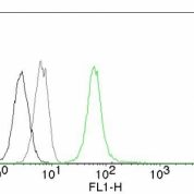
Tested Applications: Flow, IF, WB, IHC.
Clonality: Monoclonal
Anti-Vimentin (Mesenchymal Cell Marker) Antibody Clone: VM452
Clone VM452 Host and Isotype: Mouse IgG1
Anti-Human, Cow, Dog, Cat, Pig, Goat and Chicken. Does not react with mouse and rat. Vimentin (Mesenchymal Cell Marker) Positive Control Sample: Raji, Jurkat or HeLa cells. Sarcomas or Melanomas.
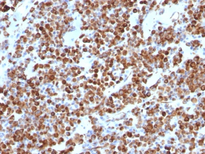
Cellular Localization of Antibody VM452 Staining: Cytoplasmic
Buffer and Stabilizer: 10mM PBS with 0.05% BSA & 0.05% azide. Also available without BSA & Azide.
Antibody Concentration: 200 ug/ml
Antibody Purification Method:Protein A/G purified from Bioreactor concentrate.
Immunogen: Recombinant full-length human vimentin protein
Storage Conditions: Store at 2 to 8 C (refrigerate). Stable for 24 months when properly stored.
Vimentin (Mesenchymal Cell Marker) Previously Observed Antibody Staining Patterns
Observed Subcellular, Organelle Specific Staining Data:
Variations in Vimentin Anti-Human antibody staining intensity in immunohistochemistry on tissue sections are present across different anatomical locations. An intense signal was observed in lymphoid tissue in appendix, hematopoietic cells in the bone marrow, adipocytes in breast, endothelial cells in the cerebral cortex and colon, glandular cells in the epididymis and fallopian tube, cells in the glomeruli in kidney, macrophages in lung, pneumocytes in lung, germinal center cells in the lymph node, non-germinal center cells in the lymph node, ovarian stroma cells in the ovary, glandular cells in the seminal vesicle, fibroblasts in skin, Langerhans in skin, melanocytes in skin, adipocytes in mesenchymal tissue, fibroblasts in mesenchymal tissue, peripheral nerve in mesenchymal tissue, cells in the red pulp in spleen, cells in the seminiferous ducts in testis, Leydig cells in the testis, glandular cells in the thyroid gland, germinal center cells in the tonsil and non-germinal center cells in the tonsil. More moderate antibody staining intensity was present in lymphoid tissue in appendix, hematopoietic cells in the bone marrow, adipocytes in breast, endothelial cells in the cerebral cortex and colon, glandular cells in the epididymis and fallopian tube, cells in the glomeruli in kidney, macrophages in lung, pneumocytes in lung, germinal center cells in the lymph node, non-germinal center cells in the lymph node, ovarian stroma cells in the ovary, glandular cells in the seminal vesicle, fibroblasts in skin, Langerhans in skin, melanocytes in skin, adipocytes in mesenchymal tissue, fibroblasts in mesenchymal tissue, peripheral nerve in mesenchymal tissue, cells in the red pulp in spleen, cells in the seminiferous ducts in testis, Leydig cells in the testis, glandular cells in the thyroid gland, germinal center cells in the tonsil and non-germinal center cells in the tonsil. Low, but measureable presence of Vimentin Anti-Human could be seen inglandular cells in the adrenal gland, respiratory epithelial cells in the bronchus, glial cells in the cerebral cortex, cells in the endometrial stroma in endometrium, glial cells in the hippocampus, bile duct cells in the liver, respiratory epithelial cells in the nasopharynx, follicle cells in the ovary, decidual cells in the placenta and squamous epithelial cells in the vagina. We were unable to detect Vimentin Anti-Human in other tissues. Disease states, inflammation, and other physiological changes can have a substantial impact on antibody staining patterns. These measurements were all taken in tissues deemed normal or from patients without known disease.Observed Antibody Staining Data By Tissue Type:
Tissues from cancer patients, for instance, have their own distinct pattern of Vimentin Anti-Human expression as measured by anti-Vimentin Anti-Human antibody immunohistochemical staining. The average level of expression by tumor is summarized in the table below. The variability row represents patient to patient variability in IHC staining.| Sample Type | breast cancer | carcinoid | cervical cancer | colorectal cancer | endometrial cancer | glioma | head and neck cancer | liver cancer | lung cancer | lymphoma | melanoma | ovarian cancer | pancreatic cancer | prostate cancer | renal cancer | skin cancer | stomach cancer | testicular cancer | thyroid cancer | urothelial cancer |
|---|---|---|---|---|---|---|---|---|---|---|---|---|---|---|---|---|---|---|---|---|
| Signal Intensity | - | - | + | - | ++ | - | ++ | ++ | ++ | +++ | +++ | + | ++ | - | +++ | ++ | ++ | ++ | +++ | ++ |
| VIM Variability | + | + | ++ | ++ | ++ | ++ | ++ | ++ | + | ++ | + | ++ | + | ++ | + | ++ | + | ++ | + | ++ |
Limitations and Warranty
enQuire Bio's product, Vimentin (Mesenchymal Cell Marker) MonoSpecific Antibody, is available for Research Use Only (RUO-Only). This antibody is guaranteed to work for a period of two years when properly stored.

There are no reviews yet.