
Mouse Anti-FcERI / FCER1A Antibody Product Attributes
Species: Mouse
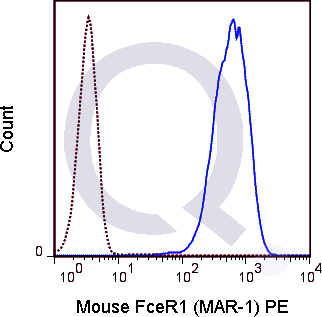
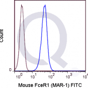
Tested Applications: Flow Cytometry.
Application Notes: See Product Datasheet for Recommended Dilution Range. Requires Experimental Optimization
Clonality: Monoclonal Antibody
Anti-FcERI / FCER1A Antibody Clone: 42795
Clone 42795 Host and Isotype: Armenian Hamster IgG
Buffer and Stabilizer: 10 mM NaH2PO4, 150 mM NaCl, 0.09% NaN3, 0.1% gelatin, pH7.2
Antibody Concentration: 0.5 mg/mL
Storage Conditions: 2-8C protected from light. Stable for 12 Months. Do Not Freeze.
FcERI / FCER1A Previously Observed Antibody Staining Patterns
Observed Antibody Staining Data By Tissue Type:
Variations in FcERI / FCER1A antibody staining intensity in immunohistochemistry on tissue sections are present across different anatomical locations. Low, but measureable presence of FcERI / FCER1A could be seen in cells in the red pulp in spleen, endothelial cells in the colon, epidermal cells in the skin, glandular cells in the adrenal gland, appendix, colon, duodenum, fallopian tube, rectum, seminal vesicle and thyroid gland, hematopoietic cells in the bone marrow, Leydig cells in the testis, melanocytes in skin, myocytes in heart muscle and skeletal muscle, myoepithelial cells in the breast, neuronal cells in the hippocampus, respiratory epithelial cells in the nasopharynx, trophoblastic cells in the placenta and urothelial cells in the urinary bladder. We were unable to detect FcERI / FCER1A in other tissues. Disease states, inflammation, and other physiological changes can have a substantial impact on antibody staining patterns. These measurements were all taken in tissues deemed normal or from patients without known disease.Observed Antibody Staining Data By Tissue Disease Status:
Tissues from cancer patients, for instance, have their own distinct pattern of FcERI / FCER1A expression as measured by anti-FcERI / FCER1A antibody immunohistochemical staining. The average level of expression by tumor is summarized in the table below. The variability row represents patient to patient variability in IHC staining.| Sample Type | breast cancer | carcinoid | cervical cancer | colorectal cancer | endometrial cancer | glioma | head and neck cancer | liver cancer | lung cancer | lymphoma | melanoma | ovarian cancer | pancreatic cancer | prostate cancer | renal cancer | skin cancer | stomach cancer | testicular cancer | thyroid cancer | urothelial cancer |
|---|---|---|---|---|---|---|---|---|---|---|---|---|---|---|---|---|---|---|---|---|
| Signal Intensity | - | - | - | - | - | - | - | + | - | - | - | - | - | - | - | - | - | - | - | - |
| FCER1A Variability | ++ | ++ | + | ++ | + | ++ | + | ++ | + | + | + | + | + | + | + | + | + | + | ++ | + |
Selected References
Mukai K, BenBarak MJ, Tachibana M, Nishida K, Karasuyama H, Taniuchi I, and Galli SJ. 2012. Blood. 120: 76-85. (Flow cytometry)Smith KA, Harcus Y, Garbi N, Hammerling GJ, MacDonald AS, and Maizels RM. 2012. Infect. Immun. 80: 3481-3489. (in vivo depletion)Larson D, Hubner MP, Torrero MN, Morris CP, Brankin A, Swierczewski BE, Davies SJ, Vonakis BM, and Mitre E. 2012. J. Immunol. 188: 4188-4199. (in vitro activation)Khodoun M, Krishnamurthy D, Strait R, Kucuk Y, and Finkelman F. 2011. J. Immunol. 186: 151.4. (in vitro depletion)
Limitations and Warranty
enQuire Bio's Mouse Anti-FCER1A Monoclonal is available for Research Use Only. This antibody is guaranteed to work for a period of two years when properly stored.

There are no reviews yet.