
 PDF Datasheet
Human, Mouse, Rat, and Pig Anti-Bcl-X Antibody Product Attributes
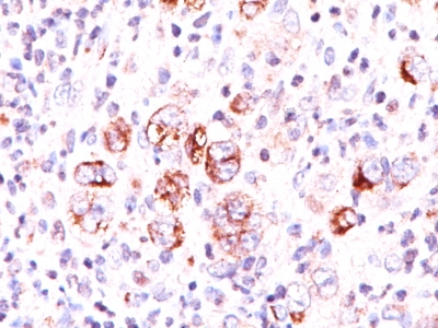
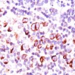
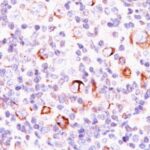
Bcl-X Previously Observed Antibody Staining Patterns
Observed Subcellular, Organelle Specific Staining Data:
Anti-BCL2L1 antibody staining is expected to be primarily localized to the mitochondria.
Observed Antibody Staining Data By Tissue Type:
Variations in Bcl-X antibody staining intensity in immunohistochemistry on tissue sections are present across different anatomical locations. Low, but measureable presence of Bcl-X could be seen in cells in the granular layer in cerebellum, chondrocytes in mesenchymal tissue, exocrine glandular cells in the pancreas, glandular cells in the colon, duodenum, endometrium, epididymis, rectum, salivary gland and thyroid gland, hepatocytes in liver, lymphoid tissue in appendix, myocytes in skeletal muscle, myoepithelial cells in the breast, neuronal cells in the caudate nucleus, non-germinal center cells in the tonsil, peripheral nerve/ganglion in colon, pneumocytes in lung, Purkinje cells in the cerebellum and squamous epithelial cells in the esophagus. We were unable to detect Bcl-X in other tissues. Disease states, inflammation, and other physiological changes can have a substantial impact on antibody staining patterns. These measurements were all taken in tissues deemed normal or from patients without known disease.
Observed Antibody Staining Data By Tissue Disease Status:
Tissues from cancer patients, for instance, have their own distinct pattern of Bcl-X expression as measured by anti-Bcl-X antibody immunohistochemical staining. The average level of expression by tumor is summarized in the table below. The variability row represents patient to patient variability in IHC staining.
| Sample Type | breast cancer | carcinoid | cervical cancer | colorectal cancer | endometrial cancer | glioma | head and neck cancer | liver cancer | lung cancer | lymphoma | melanoma | ovarian cancer | pancreatic cancer | prostate cancer | renal cancer | skin cancer | stomach cancer | testicular cancer | thyroid cancer | urothelial cancer |
|---|---|---|---|---|---|---|---|---|---|---|---|---|---|---|---|---|---|---|---|---|
| Signal Intensity | ++ | + | – | + | – | ++ | + | + | + | – | + | + | ++ | – | + | – | + | + | + | + |
| BCL2L1 Variability | ++ | ++ | ++ | ++ | ++ | ++ | ++ | ++ | ++ | + | ++ | ++ | ++ | + | ++ | ++ | +++ | ++ | ++ | ++ |
| Bcl-X General Information | |
|---|---|
| Alternate Names | |
| Bcl-2-like 1, BCL2L1 | |
| Molecular Weight | |
| 27kDa | |
| Chromosomal Location | |
| 20q11.21 | |
| Curated Database and Bioinformatic Data | |
| Gene Symbol | BCL2L1 |
| Entrez Gene ID | 598 |
| Ensemble Gene ID | ENSG00000171552 |
| RefSeq Protein Accession(s) | NP_001182, NP_001304848, NP_001304849, NP_001304850, NP_001309168, NP_001309171, XP_011527266, XP_016883482, NP_001309169, NP_612815 |
| RefSeq mRNA Accession(s) | NM_001191, NM_001317920, NM_001322240, XM_011528964, XR_001754364, NM_001317921, NM_001322239, NM_001317919, NM_138578, NM_001322242, XM_017027993, XR_936599 NR_134257 |
| RefSeq Genomic Accession(s) | NC_000020, NG_029002, NC_018931 |
| UniProt ID(s) | Q5TE63, Q07817, A0A0S2Z3C5 |
| UniGene ID(s) | Q5TE63, Q07817, A0A0S2Z3C5 |
| HGNC ID(s) | 992 |
| Cosmic ID(s) | BCL2L1 |
| KEGG Gene ID(s) | hsa:598 |
| PharmGKB ID(s) | PA76 |
| General Description of Bcl-X. | |
| Recognizes a protein of 27kDa, identified as the Bcl-X protein. This MAb shows no cross-reaction with Bcl-2 or Bax protein. Bcl-X has two isoforms, Bcl-XL (long), a 241 amino acid protein which suppresses cell death., Bcl-XS (short), a 178 amino acid protein lacking a 63 amino acid domain which functions as a dominant inhibitor of Bcl-2. This MAb reacts with both Bcl-XS, Bcl-XL proteins. | |
Reviews
There are no reviews yet.