
.jpg)
.jpg)
.jpg)
.jpg)
.jpg)
.jpg)
%20Gel.jpg)
.jpg)
 PDF Datasheet
Human and non-human primates Anti-Beta-2 Microglobulin Antibody Product Attributes
Beta-2 Microglobulin Previously Observed Antibody Staining Patterns
Observed Subcellular, Organelle Specific Staining Data:
Anti-B2M antibody staining is expected to be primarily localized to the cytosol, golgi apparatus and plasma membrane.
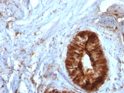
Observed Antibody Staining Data By Tissue Type:
Variations in Beta-2 Microglobulin antibody staining intensity in immunohistochemistry on tissue sections are present across different anatomical locations. An intense signal was observed in cells in the white pulp in spleen, germinal center cells in the lymph node and tonsil, hematopoietic cells in the bone marrow, macrophages in lung, non-germinal center cells in the lymph node and tonsil, pneumocytes in lung, respiratory epithelial cells in the bronchus and nasopharynx and squamous epithelial cells in the tonsil. More moderate antibody staining intensity was present in cells in the white pulp in spleen, germinal center cells in the lymph node and tonsil, hematopoietic cells in the bone marrow, macrophages in lung, non-germinal center cells in the lymph node and tonsil, pneumocytes in lung, respiratory epithelial cells in the bronchus and nasopharynx and squamous epithelial cells in the tonsil. Low, but measureable presence of Beta-2 Microglobulin could be seen inadipocytes in breast and mesenchymal tissue, bile duct cells in the liver, cells in the granular layer in cerebellum, cells in the molecular layer in cerebellum, cells in the seminiferous ducts in testis, chondrocytes in mesenchymal tissue, exocrine glandular cells in the pancreas, fibroblasts in skin and mesenchymal tissue, glandular cells in the breast and parathyroid gland, glial cells in the caudate nucleus, cerebral cortex and hippocampus, hepatocytes in liver, myocytes in heart muscle and skeletal muscle, myoepithelial cells in the breast, neuronal cells in the caudate nucleus, cerebral cortex and hippocampus, ovarian stroma cells in the ovary, peripheral nerve in mesenchymal tissue, peripheral nerve/ganglion in colon, Purkinje cells in the cerebellum, smooth muscle cells in the smooth muscle, squamous epithelial cells in the cervix and uterine and vagina. We were unable to detect Beta-2 Microglobulin in other tissues. Disease states, inflammation, and other physiological changes can have a substantial impact on antibody staining patterns. These measurements were all taken in tissues deemed normal or from patients without known disease.
Observed Antibody Staining Data By Tissue Disease Status:
Tissues from cancer patients, for instance, have their own distinct pattern of Beta-2 Microglobulin expression as measured by anti-Beta-2 Microglobulin antibody immunohistochemical staining. The average level of expression by tumor is summarized in the table below. The variability row represents patient to patient variability in IHC staining.
| Sample Type | breast cancer | carcinoid | cervical cancer | colorectal cancer | endometrial cancer | glioma | head and neck cancer | liver cancer | lung cancer | lymphoma | melanoma | ovarian cancer | pancreatic cancer | prostate cancer | renal cancer | skin cancer | stomach cancer | testicular cancer | thyroid cancer | urothelial cancer |
|---|---|---|---|---|---|---|---|---|---|---|---|---|---|---|---|---|---|---|---|---|
| Signal Intensity | + | – | + | ++ | + | – | + | ++ | – | ++ | + | + | + | + | + | – | ++ | – | ++ | + |
| B2M Variability | +++ | ++ | ++ | ++ | ++ | ++ | ++ | ++ | ++ | ++ | ++ | ++ | ++ | ++ | ++ | ++ | ++ | + | ++ | +++ |
| Beta-2 Microglobulin General Information | |
|---|---|
| Alternate Names | |
| ?2 microglobulin, B2M, B2 microglobulin | |
| Molecular Weight | |
| 12kDa | |
| Chromosomal Location | |
| 15q21-q22.2 | |
| Curated Database and Bioinformatic Data | |
| Gene Symbol | B2M |
| Entrez Gene ID | 567 |
| Ensemble Gene ID | ENSG00000166710 |
| RefSeq Protein Accession(s) | NP_004039, XP_005254606 |
| RefSeq mRNA Accession(s) | NM_004048, XM_005254549, |
| RefSeq Genomic Accession(s) | NC_018926, NC_000015, NG_187605, NG_012920 |
| UniProt ID(s) | Q9UDF4, P01884, P61769 |
| UniGene ID(s) | Q9UDF4, P01884, P61769 |
| HGNC ID(s) | 914 |
| Cosmic ID(s) | B2M |
| KEGG Gene ID(s) | hsa:567 |
| PharmGKB ID(s) | PA25207 |
| General Description of Beta-2 Microglobulin. | |
| Recognizes a protein of 12kDa, identified as beta-2 microglobulin. Major histocompatibility complex (MHC) class 1 molecules bind to antigens for presentation on the surface of cells. The proteasome is responsible for producing these antigens from the components of foreign pathogens. MHC class 1 molecules consist of an alpha heavy chain that contains three subdomains (alpha1, alpha2, alpha3), a non-covalent associating light chain, known as beta-2-Microglobulin. beta-2-Microglobulin associates with the alpha3 subdomain of the alpha heavy chain, forms an immunoglobulin domain-like structure that mediates proper folding, expression of MHC class 1 molecules. The alpha1, alpha2 domains of the alpha heavy chain form the peptide antigen-binding cleft. Mutations in the beta-2-Microglobulin gene can enhance the progression of malignant melanoma phenotypes. | |
-150x150.jpg)

-150x150.jpg)
Reviews
There are no reviews yet.