
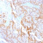
.jpg "Beta Catenin FFPE breast (ductal carcinoma)")
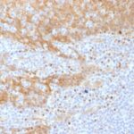
.jpg "Beta Catenin FFPE breast (lobular carcinoma)")


 PDF Datasheet
Human and Mouse Anti-beta-Catenin Antibody Product Attributes
beta-Catenin Previously Observed Antibody Staining Patterns
Observed Subcellular, Organelle Specific Staining Data:
Anti-CTNNB1 antibody staining is expected to be primarily localized to the plasma membrane.
Observed Antibody Staining Data By Tissue Type:
Variations in beta-Catenin antibody staining intensity in immunohistochemistry on tissue sections are present across different anatomical locations. An intense signal was observed in bile duct cells in the liver, cells in the seminiferous ducts in testis, cells in the tubules in kidney, epidermal cells in the skin, exocrine glandular cells in the pancreas, glandular cells in the adrenal gland, appendix, breast, cervix, uterine, colon, duodenum, endometrium, epididymis, fallopian tube, gallbladder, parathyroid gland, prostate, rectum, salivary gland, seminal vesicle, small intestine, stomach and thyroid gland, hepatocytes in liver, keratinocytes in skin, myocytes in heart muscle, myoepithelial cells in the breast, respiratory epithelial cells in the bronchus and nasopharynx, squamous epithelial cells in the cervix, uterine, esophagus, oral mucosa, tonsil and vagina, trophoblastic cells in the placenta and urothelial cells in the urinary bladder. More moderate antibody staining intensity was present in bile duct cells in the liver, cells in the seminiferous ducts in testis, cells in the tubules in kidney, epidermal cells in the skin, exocrine glandular cells in the pancreas, glandular cells in the adrenal gland, appendix, breast, cervix, uterine, colon, duodenum, endometrium, epididymis, fallopian tube, gallbladder, parathyroid gland, prostate, rectum, salivary gland, seminal vesicle, small intestine, stomach and thyroid gland, hepatocytes in liver, keratinocytes in skin, myocytes in heart muscle, myoepithelial cells in the breast, respiratory epithelial cells in the bronchus and nasopharynx, squamous epithelial cells in the cervix, uterine, esophagus, oral mucosa, tonsil and vagina, trophoblastic cells in the placenta and urothelial cells in the urinary bladder. Low, but measureable presence of beta-Catenin could be seen in cells in the endometrial stroma in endometrium, cells in the glomeruli in kidney, cells in the molecular layer in cerebellum, endothelial cells in the cerebral cortex, fibroblasts in skin, glial cells in the caudate nucleus, cerebral cortex and hippocampus, hematopoietic cells in the bone marrow, Leydig cells in the testis, neuronal cells in the caudate nucleus, cerebral cortex and hippocampus, ovarian stroma cells in the ovary and Purkinje cells in the cerebellum. We were unable to detect beta-Catenin in other tissues. Disease states, inflammation, and other physiological changes can have a substantial impact on antibody staining patterns. These measurements were all taken in tissues deemed normal or from patients without known disease.
Observed Antibody Staining Data By Tissue Disease Status:
Tissues from cancer patients, for instance, have their own distinct pattern of beta-Catenin expression as measured by anti-beta-Catenin antibody immunohistochemical staining. The average level of expression by tumor is summarized in the table below. The variability row represents patient to patient variability in IHC staining.
| Sample Type | breast cancer | carcinoid | cervical cancer | colorectal cancer | endometrial cancer | glioma | head and neck cancer | liver cancer | lung cancer | lymphoma | melanoma | ovarian cancer | pancreatic cancer | prostate cancer | renal cancer | skin cancer | stomach cancer | testicular cancer | thyroid cancer | urothelial cancer |
|---|---|---|---|---|---|---|---|---|---|---|---|---|---|---|---|---|---|---|---|---|
| Signal Intensity | +++ | ++ | +++ | +++ | +++ | + | +++ | +++ | +++ | – | +++ | +++ | +++ | +++ | ++ | +++ | ++ | + | +++ | +++ |
| CTNNB1 Variability | ++ | ++ | ++ | + | + | +++ | ++ | ++ | ++ | + | + | ++ | + | + | ++ | + | ++ | ++ | + | ++ |
| beta-Catenin General Information | |
|---|---|
| Alternate Names | |
| FCERI, FCER1A | |
| Molecular Weight | |
| 92kDa | |
| Chromosomal Location | |
| 3p22.1 | |
| Curated Database and Bioinformatic Data | |
| Gene Symbol | CTNNB1 |
| Entrez Gene ID | 1499 |
| Ensemble Gene ID | ENSG00000168036 |
| RefSeq Protein Accession(s) | XP_016861227, NP_001091679, XP_005264943, NP_001091680, XP_006713046, XP_006713047, NP_001317658, XP_006713048, NP_001895 |
| RefSeq mRNA Accession(s) | XM_006712984, XM_006712983, NM_001904, XM_017005738, NM_001098209, NM_001330729, NM_001098210, XM_006712985 |
| RefSeq Genomic Accession(s) | NC_018914, NG_013302, NC_000003 |
| UniProt ID(s) | A0A024R2Q3, P35222, B4DGU4 |
| UniGene ID(s) | A0A024R2Q3, P35222, B4DGU4 |
| HGNC ID(s) | 2514 |
| Cosmic ID(s) | CTNNB1 |
| KEGG Gene ID(s) | hsa:1499 |
| PharmGKB ID(s) | PA27013 |
| General Description of beta-Catenin. | |
| Beta-catenin associates with the cytoplasmic portion of E-cadherin, which is necessary for the function of E-cadherin as an adhesion molecule. In normal tissues, beta-catenin is localized to the membrane of epithelial cells, consistent with its role in the cell adhesion complex. In breast ductal neoplasia, beta-catenin is usually localized in cellular membranes. However, in lobular neoplasia, a marked redistribution of beta-catenin throughout the cytoplasm results in a diffuse cytoplasmic pattern. Immuno-staining of beta-catenin, E-cadherin is helps in the accurate identification of ductal, lobular neoplasms, including a distinction between low-grade ductal carcinoma in situ (DCIS), lobular carcinoma. Additionally, some rectal, gastric adenocarcinomas demonstrate diffuse cytoplasmic beta-catenin staining, a lack of membranous staining, mimicking the staining pattern observed with lobular breast carcinomas. | |

-150x150.jpg)
There are no reviews yet.