Human and Rat Anti-Calponin-1 Antibody Product Attributes
Calponin-1 Previously Observed Antibody Staining Patterns
Observed Subcellular, Organelle Specific Staining Data:
Anti-CNN1 antibody staining is expected to be primarily localized to the actin filaments. There is variability in either the signal strength or the localization of signal in actin filaments from cell to cell.
Observed Antibody Staining Data By Tissue Type:
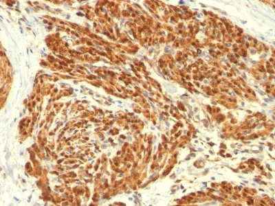
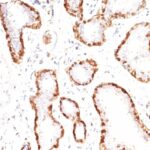
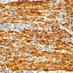
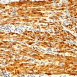
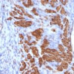
Variations in Calponin-1 antibody staining intensity in immunohistochemistry on tissue sections are present across different anatomical locations. An intense signal was observed in smooth muscle cells in the smooth muscle. More moderate antibody staining intensity was present in smooth muscle cells in the smooth muscle. Low, but measureable presence of Calponin-1 could be seen in. We were unable to detect Calponin-1 in other tissues. Disease states, inflammation, and other physiological changes can have a substantial impact on antibody staining patterns. These measurements were all taken in tissues deemed normal or from patients without known disease.
| Calponin-1 General Information | |
|---|---|
| Alternate Names | |
| Calponin 1, CNN1 | |
| Molecular Weight | |
| 34kDa | |
| Chromosomal Location | |
| 19p13.2 | |
| Curated Database and Bioinformatic Data | |
| Gene Symbol | CNN1 |
| Entrez Gene ID | 1264 |
| Ensemble Gene ID | ENSG00000130176 |
| RefSeq Protein Accession(s) | NP_001290, NP_001295270, XP_016881778, NP_001295271, XP_005259798 |
| RefSeq mRNA Accession(s) | XM_005259741, NM_001299, NM_001308342, NM_001308341, XM_017026289 |
| RefSeq Genomic Accession(s) | NC_000019, NC_018930 |
| UniProt ID(s) | Q53FP8, P51911, V9HWA5, B7Z7E1 |
| UniGene ID(s) | Q53FP8, P51911, V9HWA5, B7Z7E1 |
| HGNC ID(s) | 2155 |
| Cosmic ID(s) | CNN1 |
| KEGG Gene ID(s) | hsa:1264 |
| PharmGKB ID(s) | PA26665 |
| General Description of Calponin-1. | |
| Multiple isoelectric variants of calponin have been identified, however only two molecular weight isoforms exist; a 34kDa form, a 29kDa form. Expression of the 29kDa form, I-calponin, is primarily restricted to muscle of the urogenital tract, whereas the higher molecular weight variant has been demonstrated in vascular, visceral smooth muscle. In Western Blot, this MAb reacts with only the 34kDa form of calponin in extracts of human aortic medial smooth muscle, is unreactive with fibroblast extracts of cultivated human foreskin. Calponin is a calmodulin, F-actin, tropomyosin binding protein, which is thought to be involved in the regulation of smooth muscle contraction. Calponin expression is restricted to smooth muscle cells, has been shown to be a marker of the differentiated (contractile) phenotype of developing smooth muscle. | |


-150x150.jpg)

There are no reviews yet.