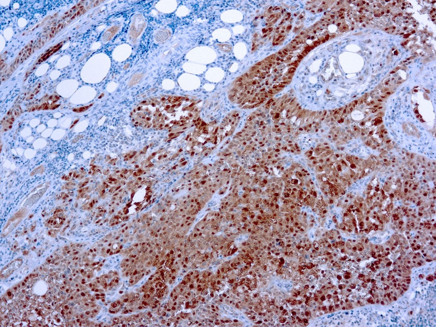
Discontinued
Antibody (Suitable for clinical applications)
Application Notes
| Specification | Recommendation |
|---|---|
| Recommended Dilution (Conc) | 1:50-1:100 |
| Pretreatment | Citrate Buffer pH 6.0 |
| Incubation Parameters | 30 min at Room Temperature |
Prior to use, inspect vial for the presence of any precipitate or other unusual physical properties. These can indicate that the antibody has degraded and is no longer suitable for patient samples. Please run positive and negative controls simultaneously with all patient samples to account and control for errors in laboratory procedure. Use of methods or materials not recommended by enQuire Bio including change to dilution range and detection system should be routinely validated by the user.
Calretinin Information for Pathologists
Summary:
Calcium binding protein structurally related to S100 and inhibin. Interpretation Nuclear and cytoplasmic staining. Uses by pathologists Differentiate (as part of a panel) epithelioid pleural mesothelioma (positive) from lung adenocarcinoma (negative, Am J Surg Pathol 2003;27:1031).
Common Uses By Pathologists:
Differentiate (as part of a panel) epithelioid pleural mesothelioma (positive) from lung adenocarcinoma (negative, Am J Surg Pathol 2003;27:1031). Differentiate (as part of a panel) epithelioid peritoneal mesothelioma (positive) from ovarian serous papillary carcinoma (usually negative, Am J Surg Pathol 2007;31:1139). Differentiate reactive mesothelial cells (positive) from carcinomas (negative) in effusion cytology (Am J Clin Pathol 2001;116:709, Cytopathology 2008;19:218), ascites fluid / peritoneal lavage (Tohoku J Exp Med 2005;206:31) or pleural biopsies (Am J Surg Pathol 2007;31:914). Differentiate (as part of a panel) mesothelioma (positive) from metastatic renal cell carcinoma (negative, Histopathology 2002;41:301). Differentiate (as part of a panel) adrenal cortical lesions (calretinin+) from metastatic clear cell renal cell carcinoma (negative, Am J Surg Pathol 2011;35:678).
| Calretinin General Information | |
|---|---|
| Alternate Names | |
| Molecular Weight | |
| 31.5 kDa | |
| Chromosomal Location | |
| [chr: CHR_HSCHR16_4_CTG3_1] [chr_start: 71363481] [chr_end: 71395224] [strand: 1]; q22.2 [chr: 16] [chr_start: 71358713] [chr_end: 71390436] [strand: 1] | |
| Curated Database and Bioinformatic Data | |
| Gene Symbol | CALB2 |
| Entrez Gene ID | 794 |
| RefSeq Protein Accession(s) | NP_001731; NP_009019 |
| RefSeq mRNA Accession(s) | NM_007088; XR_002957842; NM_001740; NR_027910; NM_007087 |
| RefSeq Genomic Accession(s) | NC_000016; NW_013171813; |
| UniProt ID(s) | P22676 |
| PharmGKB ID(s) | PA26027 |
| KEGG Gene ID(s) | hsa:794 |
| General Description of Calretinin . | |
| This antibody recognizes a 31.5 kDa protein named calretinin. Calretinin is an intracellular calcium-binding protein belonging to the troponin C superfamily characterized by a structural motif described as the EF-hand domain. The immunohistochemical detection of calretinin in developing cerebellum is restricted to the later stages indicated by weak staining from week 21 of gestation in Purkinje and basket cells and in neurons of the dentate nucleus. The intensity of staining increases as the cerebellum matures. In tumors, calretinin has been detected in mesotheliomas and some pulmonary adenocarcinomas. | |


Reviews
There are no reviews yet.