
 PDF Datasheet
Discontinued
Antibody (Suitable for clinical applications)
Application Notes
| Specification | Recommendation |
|---|---|
| Recommended Dilution (Conc) | 1:20-1:30 |
| Pretreatment | Citrate Buffer pH 6.0 |
| Incubation Parameters | 30 min at Room Temperature |
Prior to use, inspect vial for the presence of any precipitate or other unusual physical properties. These can indicate that the antibody has degraded and is no longer suitable for patient samples. Please run positive and negative controls simultaneously with all patient samples to account and control for errors in laboratory procedure. Use of methods or materials not recommended by enQuire Bio including change to dilution range and detection system should be routinely validated by the user.
CD10/CALLA Information for Pathologists
Summary:
Cell membrane metallopeptidase widely distributed in hematopoietic cells and their neoplasms. Important in diagnosis of preB-ALL (OMIM #120520). Useful in diagnosis of other entities, but must be used with caution, as staining is nonspecific
Common Uses By Pathologists:
Apical surface staining only: well differentiated carcinoma of colon, pancreas, prostate (Am J Clin Pathol 2000;113:374)
Diffuse cytoplasmic or membranous / Golgi staining pattern: adenocarcinoma (poorly differentiated), endometrial stromal sarcoma, melanoma, renal cell carcinoma, urothelial carcinoma
Acute lymphoblastic leukemia (ALL):
One of first markers to identify leukemic cells in children (hence its name)
Found on ALL cells which derive from pre-B lymphocytes
Bladder:
Present in 40%, strongly correlates with grade and stage (Diagn Pathol 2009;4:38, Am J Clin Pathol 2005;124:371)
Breast:
Marker of myoepithelial cells (Mod Pathol 2002;15:397), mammary myofibroblastoma (Virchows Arch 2007;450:727); but also rarely invasive ductal carcinoma, papilloma (J Clin Pathol 2007;60:958), benign stroma, sarcoma NOS (Diagn Pathol 2013 Jan 28;8:14)
Values may change post-chemotherapy (Indian J Cancer 2013;50:46)
Helps differentiate collagenous spherulosis (CD10+, HHF35+) from adenoid cystic carcinoma (CD10-, HHF35-, Pathol Res Pract 2012;208:405)
Ectopic prostate:
Used with PSA, PSAP to confirm diagnosis in uterus and vagina (Am J Surg Pathol 2006;30:209)
Endometrial stromal tumors:
May differentiate CD10+ endometrial stromal tumors from smooth muscle tumors, which are usually CD10- (Mod Pathol 2001;14:465), but not always (Mod Pathol 2002;15:923)
Endometriosis:
May be useful in diagnosis, except in cervix (Adv Anat Pathol 2004;11:310)
Gynecologic tumors:
Mesonephric remnants and tumors are CD10+
CD10 differentiates metastatic renal cell carcinoma (CD10+, Am J Surg Pathol 2003;27:178) from primary clear cell carcinoma (CD10-)
Hepatocellular carcinoma vs. non-hepatocellular carcinoma:
CD10+ is 52 – 68% sensitive and > 95% specific with canalicular pattern (Am J Surg Pathol 2001;25:1297, Am J Surg Pathol 2002;26:978), although another study recommends use of HepPar1, MOC31 and pCEA, but not CD10 (Mod Pathol 2002;15:1279)
Kidney
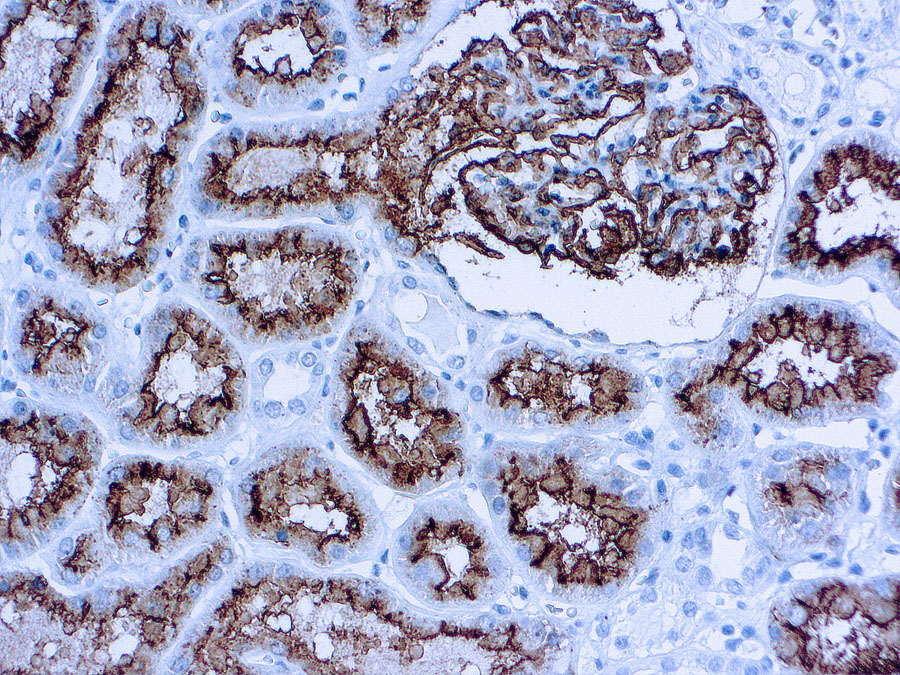
Distinguish renal cell carcinoma, clear cell type, eosinophilic variant (CD10+) from chromophobe carcinoma, eosinophilic variant or oncocytoma (both CD10-, Appl Immunohistochem Mol Morphol 2012;20:454)
Lymphoma: angioimmunoblastic T cell (AITL):
Distinguish AITL (CD10+, Mod Pathol 2011;24:993) at nodal and extranodal sites other than bone marrow from other T cell lymphomas (CD10-, Am J Surg Pathol 2004;28:54, Hum Pathol 2005;36:784), but benign T cells may also be CD10+ (Mod Pathol 2003;16:879)
Lymphoma: Burkitt:
CD10+ confirms diagnosis, but must exclude CD10+ diffuse large B cell lymphoma (Am J Clin Pathol 2012;137:665, Am J Clin Pathol 2010;133:718)
Lymphoma: diffuse large B cell:
Marker for germinal center phenotype (also HGAL, bcl6, CD38), usually considered a favorable prognostic factor (Mod Pathol 2005;18:1113, J Hematop 2009;2:187), but see Am J Clin Pathol 2001;116:183 (CD10+bcl2+ tumors have poorer survival), Virchows Arch 2004;445:545 (no difference in survival)
Lymphoma: follicular:
CD10+ may confirm diagnosis of primary (Am J Clin Pathol 2002;117:291), or secondary spread (Hum Pathol 2013;44:1328), but:
high grade follicular lymphomas and interfollicular infiltrates may be CD10- (Am J Clin Pathol 2001;115:862)
other lymphomas may be CD10+, including angioimmunoblastic T cell , Burkitt , diffuse large B cell lymphoma , mantle cell (Appl Immunohistochem Mol Morphol 2010;18:103), marginal zone (J Clin Pathol 1999;52:849), rare CD5+ CD10+ lymphomas (Am J Clin Pathol 2003;119:218, Arch Pathol Lab Med 2001;125:951)
Microvillous inclusion disease:
Strong CD10+ cytoplasmic staining in enterocytes (Am J Surg Pathol 2002;26:902, Orphanet J Rare Dis 2006 Jun 26;1:22) or colonic biopsies (Am J Surg Pathol 2010;34:970 ) vs. linear brush border staining of enterocytes and negative colonic staining in normals
Pancreas:
Confirm diagnosis of solid and papillary neoplasm (CD10+, Am J Surg Pathol 2000;24:1361)
Differentiates mucinous cystic neoplasms (CD10+ / CK20+) from intraductal papillary mucinous neoplasm of branch duct type (CD10- / CK20-, Pancreas 2009;38:558)
Skin tumors:
Staining patterns may differentiate basal cell carcinoma-epithelial staining, trichoblastoma-peritumoral stromal staining (Int J Dermatol 2009;48:713), squamous cell carcinoma-strong stromal staining (Iran J Med Sci 2013;38:100)
Differentiates atypical fibroxanthoma (diffuse membranocytoplasmic staining) from spindle cell melanoma and sarcomatoid squamous cell carcinoma (CD10-, J Cutan Pathol 2010;37:744)
Vascular tumors:
Differentiates metastatic renal cell carcinoma (CD10+, Mod Pathol 2005;18:788) from hemangioblastoma (usually CD10-, rarely focal staining-Diagn Pathol 2012 Apr 12;7:39)
Epithelioid hemangioendothelioma can have strong / diffuse CD10+ and mimic metastatic renal cell carcinoma (Arch Pathol Lab Med 2009;133:1965)
| CD10/CALLA General Information | |
|---|---|
| Alternate Names | |
| Molecular Weight | |
| 85.5 kDa | |
| Chromosomal Location | |
| q25.2 [chr: 3] [chr_start: 155024124] [chr_end: 155183729] [strand: 1] | |
| Curated Database and Bioinformatic Data | |
| Gene Symbol | MME |
| Entrez Gene ID | 4311 |
| RefSeq Protein Accession(s) | XP_011511157; NP_009219; NP_000893; XP_011511158; NP_009218; NP_009220; XP_006713709; XP_011511159; XP_006713710 |
| RefSeq mRNA Accession(s) | XM_006713647; NM_001354642; NM_001354643; NM_007289; XM_011512856; NM_007287; NM_007288; XM_011512857; NM_000902; NM_001354644 |
| RefSeq Genomic Accession(s) | NG_051105; NC_000003 |
| UniProt ID(s) | P08473 |
| PharmGKB ID(s) | PA30864 |
| KEGG Gene ID(s) | hsa:4311 |
| Associated Diseases (KEGG IDs) | Charcot-Marie-Tooth disease 2T (CMT2T) [MIM:617017]: An axonal form of Charcot-Marie-Tooth disease, a disorder of the peripheral nervous system, characterized by progressive weakness and atrophy, initially of the peroneal muscles and later of the distal muscles of the arms. Charcot-Marie-Tooth disease is classified in two main groups on the basis of electrophysiologic properties and histopathology: primary peripheral demyelinating neuropathies (designated CMT1 when they are dominantly inherited) and primary peripheral axonal neuropathies (CMT2). Neuropathies of the CMT2 group are characterized by signs of axonal degeneration in the absence of obvious myelin alterations, normal or slightly reduced nerve conduction velocities, and progressive distal muscle weakness and atrophy. {ECO:0000269|PubMed:26991897, ECO:0000269|PubMed:27588448}. The disease is caused by mutations affecting the gene represented in this entry.; Spinocerebellar ataxia 43 (SCA43) [MIM:617018]: A form of spinocerebellar ataxia, a clinically and genetically heterogeneous group of cerebellar disorders. Patients show progressive incoordination of gait and often poor coordination of hands, speech and eye movements, due to degeneration of the cerebellum with variable involvement of the brainstem and spinal cord. SCA43 is a slowly progressive, autosomal dominant form. {ECO:0000269|PubMed:27583304}. The disease is caused by mutations affecting the gene represented in this entry. |
| General Description of CD10/CALLA . | |
| This antibody is specific to human CD10 antigen of 100 kDa, also known as common acute lymphocytic leukemia antigen (CALLA). CD10 antigen has been identified on the surface of early lymphoid progenitor cells, immature B cells within adult bone marrow and germinal center B cells within lymphoid tissue. | |
Reviews
There are no reviews yet.