
![Anti-CD252 / OX40-L Antibody [RM134L] - Image 3](https://cdn-enquirebio.pressidium.com/wp-content/uploads/2017/10/enQuire-Bio-QAB75-B-100ug-anti-CD252-OX-40L-TNFSF4-antibody-10.png)
 PDF Datasheet
Mouse Anti-CD252 / OX-40L / TNFSF4 Antibody Product Attributes
CD252 / OX-40L / TNFSF4 Previously Observed Antibody Staining Patterns
Observed Subcellular, Organelle Specific Staining Data:
Staining with anti-CD252 / OX-40L / TNFSF4 antibody reveals CD252 / OX-40L / TNFSF4 expression is expected to be primarily localized to the nucleus and vesicles.
Observed Antibody Staining Data By Tissue Type:
Variations in CD252 / OX-40L / TNFSF4 antibody staining intensity in immunohistochemistry on tissue sections are present across different anatomical locations. An intense signal was observed in glandular cells in the appendix, colon and duodenum, squamous epithelial cells in the esophagus, myocytes in heart muscle, squamous epithelial cells in the oral mucosa, glandular cells in the rectum and salivary gland, epidermal cells in the skin, glandular cells in the small intestine, smooth muscle cells in the smooth muscle and cells in the seminiferous ducts in testis. More moderate antibody staining intensity was present in glandular cells in the appendix, colon and duodenum, squamous epithelial cells in the esophagus, myocytes in heart muscle, squamous epithelial cells in the oral mucosa, glandular cells in the rectum and salivary gland, epidermal cells in the skin, glandular cells in the small intestine, smooth muscle cells in the smooth muscle and cells in the seminiferous ducts in testis. Low, but measureable presence of CD252 / OX-40L / TNFSF4 could be seen inlymphoid tissue in appendix, hematopoietic cells in the bone marrow, myoepithelial cells in the breast, neuronal cells in the caudate nucleus, endothelial cells in the cerebral cortex, neuronal cells in the cerebral cortex, neuropil in cerebral cortex, endothelial cells in the colon, neuronal cells in the hippocampus, cells in the glomeruli in kidney, bile duct cells in the liver, macrophages in lung, pneumocytes in lung, germinal center cells in the lymph node, follicle cells in the ovary, exocrine glandular cells in the pancreas, glandular cells in the prostate and urothelial cells in the urinary bladder. We were unable to detect CD252 / OX-40L / TNFSF4 in other tissues. Disease states, inflammation, and other physiological changes can have a substantial impact on antibody staining patterns. These measurements were all taken in tissues deemed normal or from patients without known disease.
Observed Antibody Staining Data By Tissue Disease Status:
Tissues from cancer patients, for instance, have their own distinct pattern of CD252 / OX-40L / TNFSF4 expression as measured by anti-CD252 / OX-40L / TNFSF4 antibody immunohistochemical staining. The average level of expression by tumor is summarized in the table below. The variability row represents patient to patient variability in IHC staining.
| Sample Type | breast cancer | carcinoid | cervical cancer | colorectal cancer | endometrial cancer | glioma | head and neck cancer | liver cancer | lung cancer | lymphoma | melanoma | ovarian cancer | pancreatic cancer | prostate cancer | renal cancer | skin cancer | stomach cancer | testicular cancer | thyroid cancer | urothelial cancer |
|---|---|---|---|---|---|---|---|---|---|---|---|---|---|---|---|---|---|---|---|---|
| Signal Intensity | – | – | – | + | + | + | – | ++ | – | – | – | – | + | ++ | – | – | + | – | + | – |
| TNFSF4 Variability | ++ | + | + | ++ | ++ | ++ | ++ | ++ | ++ | + | ++ | ++ | ++ | ++ | ++ | ++ | ++ | ++ | ++ | + |
| CD252 / OX-40L / TNFSF4 General Information | |
|---|---|
| Alternate Names | |
| OX-4L, TNFSF4, CD252, GP34, OX-4L, TNLG2B, CD134L, CD252, TXGP1, OX4OL | |
| Curated Database and Bioinformatic Data | |
| Gene Symbol | Tnfsf4 |
| Entrez Gene ID | 22164 |
| Ensemble Gene ID | ENSMUSG00000026700 |
| RefSeq Protein Accession(s) | NP_033478 |
| RefSeq mRNA Accession(s) | NM_009452 |
| RefSeq Genomic Accession(s) | NC_000067 |
| UniProt ID(s) | B6DXE3, P43488 |
| UniGene ID(s) | B6DXE3, P43488 |
| Cosmic ID(s) | Tnfsf4 |
| KEGG Gene ID(s) | mmu:22164 |
| General Description of CD252 / OX-40L / TNFSF4. | |
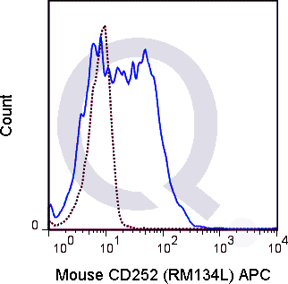
| The RM134L antibody recognizes CD252, also known as OX-40 Ligand or CD134 Ligand, a member of the TNF superfamily that is present on the surface of antigen presenting cells and activated B lymphocytes. The OX-40 Ligand interacts with OX-40 (CD134) which is expressed primarily on activated T cells. This costimulatory interaction leads to increased proliferation and IL-2 production responses of activated T cells, and at the same time enhances proliferation and immunoglobulin secretion by activated B cells.The RM134L antibody is useful for flow cytometric detection of CD252 on stimulated mouse splenocytes. It has also been reported to block the costimulatory activity of OX-40 Ligand. Please choose the appropriate format for each application. | |
There are no reviews yet.