Human Anti-CD43 Antibody Product Attributes
CD43 Previously Observed Antibody Staining Patterns
Observed Antibody Staining Data By Tissue Type:
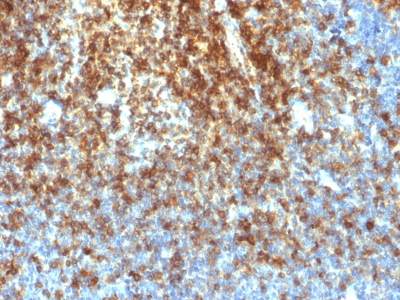
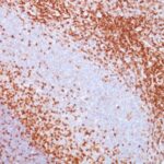
Variations in CD43 antibody staining intensity in immunohistochemistry on tissue sections are present across different anatomical locations. An intense signal was observed in lymphoid tissue in appendix, hematopoietic cells in the bone marrow, non-germinal center cells in the lymph node, cells in the red pulp in spleen and non-germinal center cells in the tonsil. More moderate antibody staining intensity was present in lymphoid tissue in appendix, hematopoietic cells in the bone marrow, non-germinal center cells in the lymph node, cells in the red pulp in spleen and non-germinal center cells in the tonsil. Low, but measureable presence of CD43 could be seen ingerminal center cells in the lymph node, tonsil. We were unable to detect CD43 in other tissues. Disease states, inflammation, and other physiological changes can have a substantial impact on antibody staining patterns. These measurements were all taken in tissues deemed normal or from patients without known disease.
| CD43 General Information | |
|---|---|
| Alternate Names | |
| Sialophorin, CD43, Leukosialin | |
| Molecular Weight | |
| 95, 115, or 135kDa | |
| Chromosomal Location | |
| 16p11.2 | |
| Curated Database and Bioinformatic Data | |
| Gene Symbol | SPN |
| Entrez Gene ID | 6693 |
| Ensemble Gene ID | ENSG00000197471 |
| RefSeq Protein Accession(s) | NP_001025459, NP_003114 |
| RefSeq mRNA Accession(s) | NM_001030288, NM_003123 |
| RefSeq Genomic Accession(s) | NC_000016, NC_018927 |
| UniProt ID(s) | P16150, A0A024R629 |
| UniGene ID(s) | P16150, A0A024R629 |
| HGNC ID(s) | 11249 |
| Cosmic ID(s) | SPN |
| KEGG Gene ID(s) | hsa:6693 |
| PharmGKB ID(s) | PA36079 |
| General Description of CD43. | |
| This MAb recognizes a cell surface glycoprotein of 95/115/135kDa (depending upon the extent of glycosylation), identified as CD43. 70-90% of T-cell lymphomas and from 22-37% of B-cell lymphomas express CD43. No reactivity has been observed with reactive B-cells. So a B-lineage population that co-expresses CD43 is highly likely to be a malignant lymphoma, especially a low-grade lymphoma, rather than a reactive B-cell population. When CD43 antibody is used in combination with anti-CD20, effective immunophenotyping of the lymphomas in formalin-fixed tissues can be obtained. Co-staining of a lymphoid infiltrate with anti-CD20 and anti-CD43 argues against a reactive process and favors a diagnosis of lymphoma. | |



There are no reviews yet.