

 PDF Datasheet
Human and Rat (-) Anti-CD57 / B3GAT1 Antibody Product Attributes
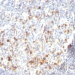
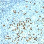
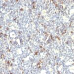
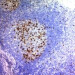
CD57 / B3GAT1 Previously Observed Antibody Staining Patterns
Observed Subcellular, Organelle Specific Staining Data:
Anti-B3GAT1 antibody staining is expected to be primarily localized to the vesicles.
Observed Antibody Staining Data By Tissue Type:
Variations in CD57 / B3GAT1 antibody staining intensity in immunohistochemistry on tissue sections are present across different anatomical locations. An intense signal was observed in glial cells in the caudate nucleus and cerebral cortex, neuronal cells in the cerebral cortex, neuropil in cerebral cortex and peripheral nerve in mesenchymal tissue. More moderate antibody staining intensity was present in glial cells in the caudate nucleus and cerebral cortex, neuronal cells in the cerebral cortex, neuropil in cerebral cortex and peripheral nerve in mesenchymal tissue. Low, but measureable presence of CD57 / B3GAT1 could be seen in cells in the red pulp in spleen, cells in the tubules in kidney, cells in the white pulp in spleen, germinal center cells in the lymph node and tonsil, glandular cells in the adrenal gland, appendix, colon, duodenum, prostate, rectum, small intestine and stomach, non-germinal center cells in the lymph node and tonsil and Purkinje cells in the cerebellum. We were unable to detect CD57 / B3GAT1 in other tissues. Disease states, inflammation, and other physiological changes can have a substantial impact on antibody staining patterns. These measurements were all taken in tissues deemed normal or from patients without known disease.
Observed Antibody Staining Data By Tissue Disease Status:
Tissues from cancer patients, for instance, have their own distinct pattern of CD57 / B3GAT1 expression as measured by anti-CD57 / B3GAT1 antibody immunohistochemical staining. The average level of expression by tumor is summarized in the table below. The variability row represents patient to patient variability in IHC staining.
| Sample Type | breast cancer | carcinoid | cervical cancer | colorectal cancer | endometrial cancer | glioma | head and neck cancer | liver cancer | lung cancer | lymphoma | melanoma | ovarian cancer | pancreatic cancer | prostate cancer | renal cancer | skin cancer | stomach cancer | testicular cancer | thyroid cancer | urothelial cancer |
|---|---|---|---|---|---|---|---|---|---|---|---|---|---|---|---|---|---|---|---|---|
| Signal Intensity | – | + | – | – | – | ++ | – | – | – | – | – | – | – | ++ | – | – | – | – | – | – |
| B3GAT1 Variability | + | +++ | + | + | + | ++ | + | + | + | + | + | + | + | + | + | + | + | + | + | + |
| CD57 / B3GAT1 General Information | |
|---|---|
| Alternate Names | |
| Galactosylgalactosylxylosylprotein 3-beta-glucuronosyltransferase 1, B3GAT1, | |
| Molecular Weight | |
| ~110kDa | |
| Chromosomal Location | |
| 11q25 | |
| Curated Database and Bioinformatic Data | |
| Gene Symbol | B3GAT1 |
| Entrez Gene ID | 27087 |
| Ensemble Gene ID | ENSG00000109956 |
| RefSeq Protein Accession(s) | XP_016873039, XP_005271563, NP_473366, XP_016873041, XP_011541053, XP_016873040, XP_011541055, NP_061114 |
| RefSeq mRNA Accession(s) | NM_018644, XM_005271506, XM_017017550, XM_011542753, XM_011542751, XM_017017551, NM_054025, XM_017017552 |
| RefSeq Genomic Accession(s) | NC_018922, NC_000011 |
| UniProt ID(s) | Q9P2W7 |
| UniGene ID(s) | Q9P2W7 |
| HGNC ID(s) | 921 |
| Cosmic ID(s) | B3GAT1 |
| KEGG Gene ID(s) | hsa:27087 |
| PharmGKB ID(s) | PA25215 |
| General Description of CD57 / B3GAT1. | |
| Anti-CD57 marks a subset of lymphocytes known as natural killer (NK) cells. Follicular center cell lymphomas often contain many NK cells within the neoplastic follicles. Anti-CD57 also stains neuroendocrine cells, their derived tumors, including carcinoid tumor, medulloblastoma. Anti-CD57 can also be useful in separating type B3 thymoma from thymic carcinoma when combined with a panel that includes antibodies against GLUT1, CD5,, CEA. | |
There are no reviews yet.