

 PDF Datasheet
Human Anti-CD63 Antibody Product Attributes
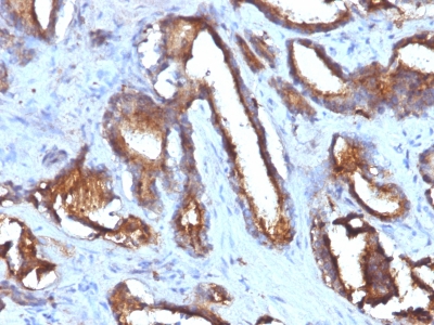
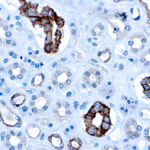
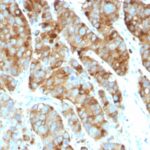
CD63 Previously Observed Antibody Staining Patterns
Observed Subcellular, Organelle Specific Staining Data:
Anti-CD63 antibody staining is expected to be primarily localized to the vesicles.
Observed Antibody Staining Data By Tissue Type:
Variations in CD63 antibody staining intensity in immunohistochemistry on tissue sections are present across different anatomical locations. Low, but measureable presence of CD63 could be seen in cells in the seminiferous ducts in testis, cells in the tubules in kidney, decidual cells in the placenta, endothelial cells in the colon, exocrine glandular cells in the pancreas, glandular cells in the adrenal gland, appendix, colon, duodenum, endometrium, parathyroid gland and small intestine, hepatocytes in liver, islets of Langerhans in pancreas, melanocytes in skin, myocytes in heart muscle, non-germinal center cells in the lymph node and tonsil, peripheral nerve/ganglion in colon, respiratory epithelial cells in the bronchus and squamous epithelial cells in the tonsil. We were unable to detect CD63 in other tissues. Disease states, inflammation, and other physiological changes can have a substantial impact on antibody staining patterns. These measurements were all taken in tissues deemed normal or from patients without known disease.
Observed Antibody Staining Data By Tissue Disease Status:
Tissues from cancer patients, for instance, have their own distinct pattern of CD63 expression as measured by anti-CD63 antibody immunohistochemical staining. The average level of expression by tumor is summarized in the table below. The variability row represents patient to patient variability in IHC staining.
| Sample Type | breast cancer | carcinoid | cervical cancer | colorectal cancer | endometrial cancer | glioma | head and neck cancer | liver cancer | lung cancer | lymphoma | melanoma | ovarian cancer | pancreatic cancer | prostate cancer | renal cancer | skin cancer | stomach cancer | testicular cancer | thyroid cancer | urothelial cancer |
|---|---|---|---|---|---|---|---|---|---|---|---|---|---|---|---|---|---|---|---|---|
| Signal Intensity | – | – | – | – | + | – | – | – | + | – | +++ | + | + | + | + | – | – | – | + | – |
| CD63 Variability | ++ | ++ | ++ | ++ | ++ | + | + | ++ | +++ | ++ | ++ | ++ | ++ | ++ | ++ | + | ++ | ++ | + | ++ |
| CD63 General Information | |
|---|---|
| Alternate Names | |
| CD63 antigen, CD63, Cluster of Differentiation 63 | |
| Molecular Weight | |
| 26kDa (core protein); 30-60kDa (glycosylated) | |
| Chromosomal Location | |
| 12q13.2 | |
| Curated Database and Bioinformatic Data | |
| Gene Symbol | CD63 |
| Entrez Gene ID | 967 |
| Ensemble Gene ID | ENSG00000135404 |
| RefSeq Protein Accession(s) | NP_001244320, NP_001254627, NP_001771, NP_001244321, NP_001244329, NP_001244319, NP_001244330, NP_001244318 |
| RefSeq mRNA Accession(s) | NM_001780, NM_001040034, NM_001257401, NM_001267698, NM_001257392, NM_001257391, NM_001257400, NM_001257389, NM_001257390 |
| RefSeq Genomic Accession(s) | NG_008347, NC_018923, NC_000012 |
| UniProt ID(s) | P08962, A0A024RB05 |
| UniGene ID(s) | P08962, A0A024RB05 |
| HGNC ID(s) | 1692 |
| Cosmic ID(s) | CD63 |
| KEGG Gene ID(s) | hsa:967 |
| PharmGKB ID(s) | PA26231 |
| General Description of CD63. | |
| This MAb recognizes protein of 26kDa-60kDa, which is identified as CD63. The tetraspanins are integral membrane proteins expressed on cell surface, granular membranes of hematopoietic cells, are components of multi-molecular complexes with specific integrins. The tetraspanin CD63 is a lysosomal membrane glycoprotein that translocates to the plasma membrane after platelet activation. CD63 is expressed on activated platelets, monocytes, macrophages,, is weakly expressed on granulocytes, T cell, B cells. It is located on the basophilic granule membranes, on the plasma membranes of lymphocytes, granulocytes. CD63 is a member of the TM4 superfamily of leukocyte glycoproteins that includes CD9, CD37, CD53, which contain four transmembrane regions. CD63 may play a role in phagocytic, intracellular lysosome-phagosome fusion events. CD63 deficiency is associated with Hermansky-Pudlak syndrome, is strongly expressed during the early stages of melanoma progression. | |
Reviews
There are no reviews yet.