
 PDF Datasheet
Human and Mouse Anti-CD74 Antibody Product Attributes
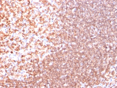
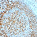
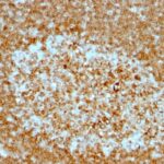
CD74 Previously Observed Antibody Staining Patterns
Observed Subcellular, Organelle Specific Staining Data:
Anti-CD74 antibody staining is expected to be primarily localized to the golgi apparatus. There is variability in either the signal strength or the localization of signal in golgi apparatus from cell to cell.
Observed Antibody Staining Data By Tissue Type:
Variations in CD74 antibody staining intensity in immunohistochemistry on tissue sections are present across different anatomical locations. Low, but measureable presence of CD74 could be seen in cells in the molecular layer in cerebellum, cells in the tubules in kidney, endothelial cells in the cerebral cortex, follicle cells in the ovary, glandular cells in the appendix, cervix, uterine, endometrium, small intestine, stomach and thyroid gland, glial cells in the caudate nucleus, cerebral cortex and hippocampus, hematopoietic cells in the bone marrow, Leydig cells in the testis, neuronal cells in the caudate nucleus, respiratory epithelial cells in the nasopharynx, squamous epithelial cells in the esophagus and trophoblastic cells in the placenta. We were unable to detect CD74 in other tissues. Disease states, inflammation, and other physiological changes can have a substantial impact on antibody staining patterns. These measurements were all taken in tissues deemed normal or from patients without known disease.
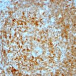
Observed Antibody Staining Data By Tissue Disease Status:
Tissues from cancer patients, for instance, have their own distinct pattern of CD74 expression as measured by anti-CD74 antibody immunohistochemical staining. The average level of expression by tumor is summarized in the table below. The variability row represents patient to patient variability in IHC staining.
| Sample Type | breast cancer | carcinoid | cervical cancer | colorectal cancer | endometrial cancer | glioma | head and neck cancer | liver cancer | lung cancer | lymphoma | melanoma | ovarian cancer | pancreatic cancer | prostate cancer | renal cancer | skin cancer | stomach cancer | testicular cancer | thyroid cancer | urothelial cancer |
|---|---|---|---|---|---|---|---|---|---|---|---|---|---|---|---|---|---|---|---|---|
| Signal Intensity | – | – | ++ | + | ++ | + | ++ | + | + | ++ | – | + | – | + | + | – | – | – | ++ | – |
| CD74 Variability | ++ | + | ++ | ++ | ++ | ++ | ++ | ++ | ++ | + | ++ | +++ | ++ | ++ | ++ | ++ | ++ | + | ++ | ++ |
| CD74 General Information | |
|---|---|
| Alternate Names | |
| CLIP, HLA class II histocompatibility antigen gamma chain, HLA-DR antigens-associated invariant chain, CD74, Cluster of Differentiation 74 | |
| Molecular Weight | |
| 33-41kDa | |
| Chromosomal Location | |
| 5q33.1 | |
| Curated Database and Bioinformatic Data | |
| Gene Symbol | CD74 |
| Entrez Gene ID | 972 |
| Ensemble Gene ID | ENSG00000019582 |
| RefSeq Protein Accession(s) | NP_001020329, NP_001020330, NP_004346, XP_016865579, XP_016865578 |
| RefSeq mRNA Accession(s) | NM_004355 NM_001025158, NM_001025159, XM_017010089, XM_017010090 |
| RefSeq Genomic Accession(s) | NG_029730, NC_018916, NC_000005 |
| UniProt ID(s) | P04233 |
| UniGene ID(s) | P04233 |
| HGNC ID(s) | 1697 |
| Cosmic ID(s) | CD74 |
| KEGG Gene ID(s) | hsa:972 |
| PharmGKB ID(s) | PA26236 |
| General Description of CD74. | |
| CD74 is a type II transmembrane protein which binds to the peptide binding groove of newly synthesized MHC class II alpha/beta heterodimers, prevents their premature association with enCanineenous polypeptides. CD74 is expressed primarily by antigen presenting cells, such as B-lymphocytes (from before the pre-B cell stage to before the plasma cell stage), macrophages,, monocytes,, many epithelial cells. Anti-CD74 stains predominantly germinal center lymphocytes, B-cell lymphomas, but rarely T-cell lymphomas. Anti-CD74 has been shown to be useful in differentiating atypical fibroxanthoma (-) from malignant fibrous histiocytoma (+). | |

There are no reviews yet.