

 PDF Datasheet
Human, Mouse, and Rat Anti-CD81 / TAPA-1 Antibody Product Attributes
CD81 / TAPA-1 Previously Observed Antibody Staining Patterns
Observed Subcellular, Organelle Specific Staining Data:
Anti-CD81 antibody staining is expected to be primarily localized to the plasma membrane.
Observed Antibody Staining Data By Tissue Type:
Variations in CD81 / TAPA-1 antibody staining intensity in immunohistochemistry on tissue sections are present across different anatomical locations. An intense signal was observed in cells in the endometrial stroma in endometrium, cells in the seminiferous ducts in testis, germinal center cells in the tonsil and ovarian stroma cells in the ovary. More moderate antibody staining intensity was present in cells in the endometrial stroma in endometrium, cells in the seminiferous ducts in testis, germinal center cells in the tonsil and ovarian stroma cells in the ovary. Low, but measureable presence of CD81 / TAPA-1 could be seen inadipocytes in mesenchymal tissue, cells in the red pulp in spleen, exocrine glandular cells in the pancreas, glandular cells in the seminal vesicle, hematopoietic cells in the bone marrow, islets of Langerhans in pancreas, keratinocytes in skin, melanocytes in skin, myocytes in heart muscle and non-germinal center cells in the tonsil. We were unable to detect CD81 / TAPA-1 in other tissues. Disease states, inflammation, and other physiological changes can have a substantial impact on antibody staining patterns. These measurements were all taken in tissues deemed normal or from patients without known disease.
Observed Antibody Staining Data By Tissue Disease Status:
Tissues from cancer patients, for instance, have their own distinct pattern of CD81 / TAPA-1 expression as measured by anti-CD81 / TAPA-1 antibody immunohistochemical staining. The average level of expression by tumor is summarized in the table below. The variability row represents patient to patient variability in IHC staining.
| Sample Type | breast cancer | carcinoid | cervical cancer | colorectal cancer | endometrial cancer | glioma | head and neck cancer | liver cancer | lung cancer | lymphoma | melanoma | ovarian cancer | pancreatic cancer | prostate cancer | renal cancer | skin cancer | stomach cancer | testicular cancer | thyroid cancer | urothelial cancer |
|---|---|---|---|---|---|---|---|---|---|---|---|---|---|---|---|---|---|---|---|---|
| Signal Intensity | – | – | – | – | – | – | – | – | – | + | – | – | – | – | – | – | – | – | – | – |
| CD81 Variability | + | + | + | + | ++ | ++ | + | ++ | + | ++ | ++ | ++ | + | + | ++ | + | + | ++ | ++ | + |
| CD81 / TAPA-1 General Information | |
|---|---|
| Alternate Names | |
| CD81 molecule, CD81, Cluster of Differentiation 81 | |
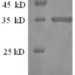
| Molecular Weight | |
| 26kDa | |
| Chromosomal Location | |
| 11p15.5 | |
| Curated Database and Bioinformatic Data | |
| Gene Symbol | CD81 |
| Entrez Gene ID | 975 |
| Ensemble Gene ID | ENSG00000110651 |
| RefSeq Protein Accession(s) | NP_004347, XP_011518794, XP_016874087, NP_001284578 |
| RefSeq mRNA Accession(s) | NM_001297649, NM_004356, XM_011520492 |
| RefSeq Genomic Accession(s) | NG_023386, NC_000011, NC_018922 |
| UniProt ID(s) | P18582, A0A024RCB7, P60033, E9PJK1 |
| UniGene ID(s) | P18582, A0A024RCB7, P60033, E9PJK1 |
| HGNC ID(s) | 1701 |
| Cosmic ID(s) | CD81 |
| KEGG Gene ID(s) | hsa:975 |
| PharmGKB ID(s) | PA26240 |
| General Description of CD81 / TAPA-1. | |
| Recognizes a protein of 26kDa, identified as CD81 (Workshop VI ;Code CD81.1)). CD81 has a very broad cellular distribution, being expressed on T- and B-lymphocytes, NK cells, thymocytes, eosinophils, fibroblasts, epithelial and endothelial cells. Neutrophils, erythrocytes and platelets are negative, while monocytes are variably positive. CD81 is a member of a family of tetraspans transmembrane proteins, including CD9, CD37, CD53, CD63, and CD82. It associates with CD19, CD21, Leu 13, and integrins on cell membrane and is involved in signal transduction in B lymphocyte development and cell adhesion. CD81 also acts as a receptor for the envelope protein E2 of chronic hepatitis C virus. Antibodies to CD81 have anti-proliferative effects on different lymphoid cell lines, particularly those derived from large cell lymphomas. | |
There are no reviews yet.