Mouse Anti-CD90.2 Antibody Product Attributes
CD90.2 Previously Observed Antibody Staining Patterns
Observed Subcellular, Organelle Specific Staining Data:
Staining with anti-CD90.2 antibody reveals CD90.2 expression is expected to be primarily localized to the nucleus and plasma membrane.
Observed Antibody Staining Data By Tissue Type:
Variations in CD90.2 antibody staining intensity in immunohistochemistry on tissue sections are present across different anatomical locations. Low, but measureable presence of CD90.2 could be seen inperipheral nerve/ganglion in colon, cells in the tubules in kidney and smooth muscle cells in the smooth muscle. We were unable to detect CD90.2 in other tissues. Disease states, inflammation, and other physiological changes can have a substantial impact on antibody staining patterns. These measurements were all taken in tissues deemed normal or from patients without known disease.
| CD90.2 General Information | |
|---|---|
| Alternate Names | |
| Thy-1.2 | |
| Curated Database and Bioinformatic Data | |
| Gene Symbol | Thy1 |
| Entrez Gene ID | 21838 |
| Ensemble Gene ID | ENSMUSG00000032011 |
| RefSeq Protein Accession(s) | NP_033408 |
| RefSeq mRNA Accession(s) | NM_009382 |
| RefSeq Genomic Accession(s) | NC_000075 |
| UniProt ID(s) | P01831 |
| UniGene ID(s) | P01831 |
| Cosmic ID(s) | Thy1 |
| KEGG Gene ID(s) | mmu:21838 |
| General Description of CD90.2. | |
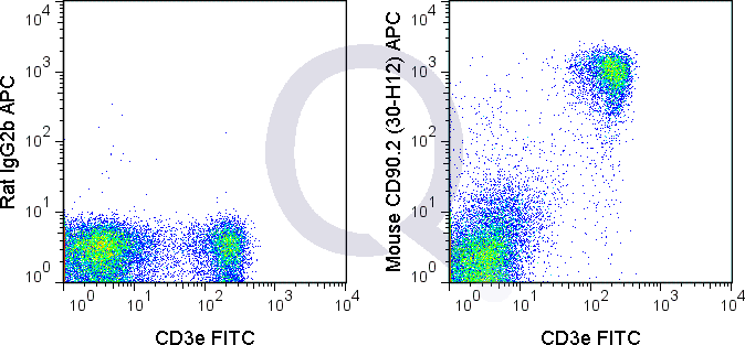
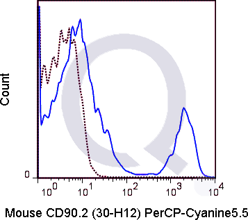
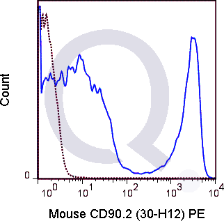
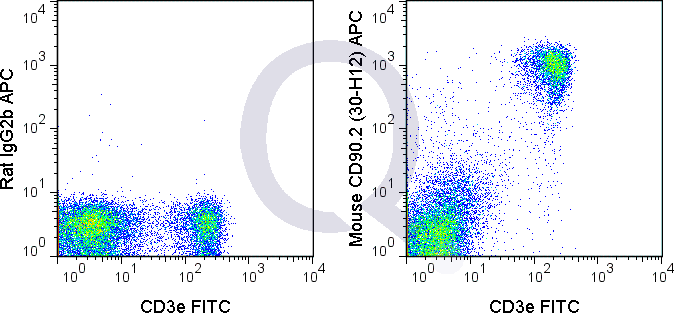
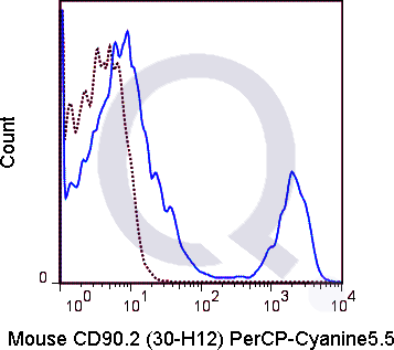
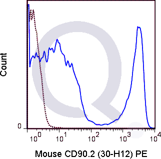
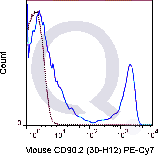
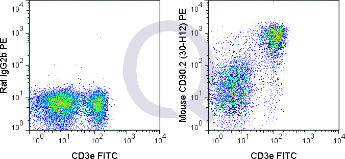
| The 30-H12 antibody reacts with mouse CD90.2 (Thy1.2). CD90.2 is a strain-specific allelic form of the GPI-linked membrane associated protein CD90 and is involved in adhesion and signal transduction. CD90.2 is expressed on thymocytes, mature T cells and neurons in mouse strains that express the CD90.2 allele (BALB/c, CBA, C3H, C57BL/6, SJL and others). 30-H12 does not react with the CD90.1 allele expressed in mouse strains such as PL and AKR. | |






There are no reviews yet.