

 PDF Datasheet
Human and Rat Anti-CD99 / MIC2 Antibody Product Attributes
Storage, Stability
Store at 2 to 8° C (refrigerate).. Stable for 24 months when properly stored.
Limitations
This antibody is available for research use only, is not approved for use in diagnostics.
Warranty
This antibody is good to the last drop. If you have any problems, call or email us immediately.
Stocked, validated by enQuire Bio, manufactured using only the highest quality standards by NeoBiotechnologies.
Search terms: 12E7; E2 antigen; MIC 2X; MIC 2Y; MIC2; Protein MIC2; Surface antigen MIC2; T-cell surface glycoprotein E2; anti-12E7 antibody; anti-E2 antigen antibody; anti-MIC 2X antibody; anti-MIC 2Y antibody; anti-MIC2 antibody; anti-Protein MIC2 antibody; anti-Surface antigen MIC2 antibody; anti-T-cell surface glycoprotein E2 antibody
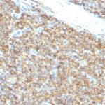
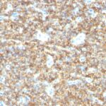
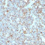
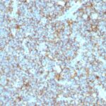
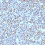
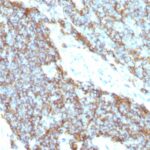
CD99 / MIC2 Previously Observed Antibody Staining Patterns
Observed Subcellular, Organelle Specific Staining Data:
Anti-CD99 antibody staining is expected to be primarily localized to the golgi apparatus.
Observed Antibody Staining Data By Tissue Type:
Variations in CD99 / MIC2 antibody staining intensity in immunohistochemistry on tissue sections are present across different anatomical locations. An intense signal was observed in cells in the endometrial stroma in endometrium, cells in the red pulp in spleen, cells in the seminiferous ducts in testis, glandular cells in the cervix, uterine and prostate, hematopoietic cells in the bone marrow, islets of Langerhans in pancreas, ovarian stroma cells in the ovary, respiratory epithelial cells in the nasopharynx and squamous epithelial cells in the esophagus. More moderate antibody staining intensity was present in cells in the endometrial stroma in endometrium, cells in the red pulp in spleen, cells in the seminiferous ducts in testis, glandular cells in the cervix, uterine and prostate, hematopoietic cells in the bone marrow, islets of Langerhans in pancreas, ovarian stroma cells in the ovary, respiratory epithelial cells in the nasopharynx and squamous epithelial cells in the esophagus. Low, but measureable presence of CD99 / MIC2 could be seen inadipocytes in mesenchymal tissue, cells in the white pulp in spleen, endothelial cells in the colon, follicle cells in the ovary, glandular cells in the breast, duodenum, endometrium, epididymis and gallbladder, macrophages in lung, non-germinal center cells in the tonsil and squamous epithelial cells in the tonsil. We were unable to detect CD99 / MIC2 in other tissues. Disease states, inflammation, and other physiological changes can have a substantial impact on antibody staining patterns. These measurements were all taken in tissues deemed normal or from patients without known disease.
Observed Antibody Staining Data By Tissue Disease Status:
Tissues from cancer patients, for instance, have their own distinct pattern of CD99 / MIC2 expression as measured by anti-CD99 / MIC2 antibody immunohistochemical staining. The average level of expression by tumor is summarized in the table below. The variability row represents patient to patient variability in IHC staining.
| Sample Type | breast cancer | carcinoid | cervical cancer | colorectal cancer | endometrial cancer | glioma | head and neck cancer | liver cancer | lung cancer | lymphoma | melanoma | ovarian cancer | pancreatic cancer | prostate cancer | renal cancer | skin cancer | stomach cancer | testicular cancer | thyroid cancer | urothelial cancer |
|---|---|---|---|---|---|---|---|---|---|---|---|---|---|---|---|---|---|---|---|---|
| Signal Intensity | – | ++ | – | + | – | +++ | ++ | – | – | + | – | – | – | – | – | – | – | – | – | – |
| CD99 Variability | ++ | ++ | + | ++ | ++ | ++ | ++ | ++ | + | ++ | ++ | + | ++ | ++ | ++ | ++ | ++ | + | ++ | ++ |
| CD99 / MIC2 General Information | |
|---|---|
| Alternate Names | |
| CD99 antigen, Cluster of differentiation 99, MIC2, single-chain type-1 glycoprotein | |
| Molecular Weight | |
| 27-32kDa | |
| Chromosomal Location | |
| Xp22.33 | |
| Curated Database and Bioinformatic Data | |
| Gene Symbol | CD99 |
| Entrez Gene ID | 4267 |
| Ensemble Gene ID | ENSG00000002586 |
| RefSeq Protein Accession(s) | NP_002405, NP_001308298, NP_001116370, NP_001308296, NP_001308297, NP_001308299 |
| RefSeq mRNA Accession(s) | NM_001321368, NM_001321369, NM_001321370, NM_001122898, NM_002414, NM_001277710, NM_001321367, NR_135623 |
| RefSeq Genomic Accession(s) | NC_000023, NC_000024, NG_009174, NC_018934 |
| UniProt ID(s) | A0A096LP69, P14209 |
| UniGene ID(s) | A0A096LP69, P14209 |
| HGNC ID(s) | 7082 |
| Cosmic ID(s) | CD99 |
| KEGG Gene ID(s) | hsa:4267 |
| PharmGKB ID(s) | PA30804 |
| General Description of CD99 / MIC2. | |
| Recognizes a sialoglycoprotein of 27-32kDa, identified as CD99, or MIC2 gene product, or E2 antigen. This antigen is expressed on the cell membrane of some lymphocytes, cortical thymocytes,, granulosa cells of the ovary. Most pancreatic islet cells, Sertoli cells of the testis,, some endothelial cells express this antigen. Mature granulocytes express very little or no CD99. MIC2 is strongly expressed on Ewing s sarcoma cells, primitive peripheral neuroectodermal tumors. This MAb shows a very similar reactivity to other CD99 MAbs (e.g. O13, 12E7, or HBA-71), is excellent for immunohistochemical staining of formalin-fixed, paraffin-embedded tissues. | |
There are no reviews yet.