
 PDF Datasheet
Human, Mouse, and Rat Anti-Cytokeratin 14 Antibody Product Attributes
Cytokeratin 14 Previously Observed Antibody Staining Patterns
Observed Subcellular, Organelle Specific Staining Data:
Anti-KRT14 antibody staining is expected to be primarily localized to the intermediate filaments. There is variability in either the signal strength or the localization of signal in intermediate filaments from cell to cell.
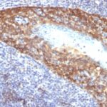
Observed Antibody Staining Data By Tissue Type:
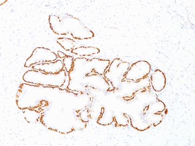
Variations in Cytokeratin 14 antibody staining intensity in immunohistochemistry on tissue sections are present across different anatomical locations. An intense signal was observed in epidermal cells in the skin, myoepithelial cells in the breast, squamous epithelial cells in the cervix and uterine, esophagus, oral mucosa, tonsil and vagina. More moderate antibody staining intensity was present in epidermal cells in the skin, myoepithelial cells in the breast, squamous epithelial cells in the cervix and uterine, esophagus, oral mucosa, tonsil and vagina. Low, but measureable presence of Cytokeratin 14 could be seen inglandular cells in the breast. We were unable to detect Cytokeratin 14 in other tissues. Disease states, inflammation, and other physiological changes can have a substantial impact on antibody staining patterns. These measurements were all taken in tissues deemed normal or from patients without known disease.
Observed Antibody Staining Data By Tissue Disease Status:
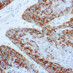
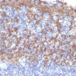
Tissues from cancer patients, for instance, have their own distinct pattern of Cytokeratin 14 expression as measured by anti-Cytokeratin 14 antibody immunohistochemical staining. The average level of expression by tumor is summarized in the table below. The variability row represents patient to patient variability in IHC staining.
| Sample Type | breast cancer | carcinoid | cervical cancer | colorectal cancer | endometrial cancer | glioma | head and neck cancer | liver cancer | lung cancer | lymphoma | melanoma | ovarian cancer | pancreatic cancer | prostate cancer | renal cancer | skin cancer | stomach cancer | testicular cancer | thyroid cancer | urothelial cancer |
|---|---|---|---|---|---|---|---|---|---|---|---|---|---|---|---|---|---|---|---|---|
| Signal Intensity | – | – | ++ | – | – | – | ++ | – | – | – | – | – | – | – | – | +++ | – | – | – | – |
| KRT14 Variability | + | + | ++ | + | + | + | ++ | + | ++ | + | + | + | + | + | + | + | + | + | + | ++ |
| Cytokeratin 14 General Information | |
|---|---|
| Alternate Names | |
| Keratin 14 | |
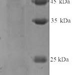
| Molecular Weight | |
| 50kDa | |
| Chromosomal Location | |
| 17q21.2 | |
| Curated Database and Bioinformatic Data | |
| Gene Symbol | KRT14 |
| Entrez Gene ID | 3861 |
| Ensemble Gene ID | ENSG00000186847 |
| RefSeq Protein Accession(s) | NP_000517 |
| RefSeq mRNA Accession(s) | NM_000526 |
| RefSeq Genomic Accession(s) | NG_008624, NC_000017, NC_018928 |
| UniProt ID(s) | P02533 |
| UniGene ID(s) | P02533 |
| HGNC ID(s) | 6416 |
| Cosmic ID(s) | KRT14 |
| KEGG Gene ID(s) | hsa:3861 |
| PharmGKB ID(s) | PA30203 |
| General Description of Cytokeratin 14. | |
| Cytokeratin 14 (CK14) belongs to the type I (or A or acidic) subfamily of low molecular weight keratins, exists in combination with keratin 5 (type II or B or basic). CK14 is found in basal cells of squamous epithelia, some glandular epithelia, myoepithelium,, mesothelial cells. Anti-CK14 is useful in differentiating squamous cell carcinomas from poorly differentiated epithelial tumors. Anti-CK14 is one of the specific basal markers for distinguishing between basal, non-basal subtypes of breast carcinomas. Anti-CK14 is also a good marker for differentiation of intraductal from invasive salivary duct carcinoma by the positive staining of basal cells surrounding the in-situ neoplasm as well as for differentiation of benign prostate from prostate carcinoma. Furthermore, this antibody has been useful in separating oncocytic tumors of the kidney from its renal mimics,, in identifying metaplastic carcinomas of the breast. | |
Reviews
There are no reviews yet.