
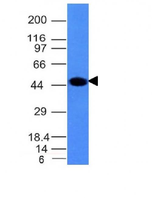
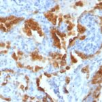
![Analysis of Mass Spec data (dashed-line) of fractions stained with Cytokeratin 8 MS-QAVA™ monoclonal antibody [Clone: H1] (solid-line), reveals that less than 11.6% of signal is attributable to non-specific binding of anti-Cytokeratin 8 [Clone H1] to targets other than KRT8 protein. Even frequently cited antibodies have much greater non-specific interactions, averaging over 30%. Data in image is from analysis in Jurkat, U202 and HeLa cells.](https://cdn-enquirebio.pressidium.com/wp-content/uploads/2017/10/enQuire-Bio-3856-MSM1-P1-anti-Cytokeratin-8-antibody.png)
 PDF Datasheet
Human, Rat, and Zebrafish Anti-Cytokeratin 8 Antibody Product Attributes
Cytoplasmic
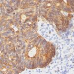
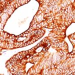
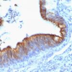
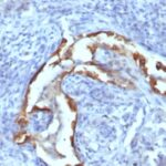
Cytokeratin 8 Previously Observed Antibody Staining Patterns
Observed Subcellular, Organelle Specific Staining Data:
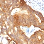
Variations in Cytokeratin 8 antibody staining intensity in immunohistochemistry on tissue sections are present across different anatomical locations. An intense signal was observed in bile duct cells in the liver, exocrine glandular cells in the pancreas, follicle cells in the ovary, glandular cells in the appendix, breast, cervix, uterine, duodenum, endometrium, epididymis, fallopian tube, gallbladder, prostate, rectum, seminal vesicle, small intestine, stomach and thyroid gland, respiratory epithelial cells in the bronchus and nasopharynx, trophoblastic cells in the placenta and urothelial cells in the urinary bladder. More moderate antibody staining intensity was present in bile duct cells in the liver, exocrine glandular cells in the pancreas, follicle cells in the ovary, glandular cells in the appendix, breast, cervix, uterine, duodenum, endometrium, epididymis, fallopian tube, gallbladder, prostate, rectum, seminal vesicle, small intestine, stomach and thyroid gland, respiratory epithelial cells in the bronchus and nasopharynx, trophoblastic cells in the placenta and urothelial cells in the urinary bladder. Low, but measureable presence of Cytokeratin 8 could be seen in. We were unable to detect Cytokeratin 8 in other tissues. Disease states, inflammation, and other physiological changes can have a substantial impact on antibody staining patterns. These measurements were all taken in tissues deemed normal or from patients without known disease.
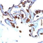
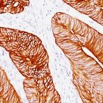
Observed Antibody Staining Data By Tissue Type:
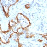
Tissues from cancer patients, for instance, have their own distinct pattern of Cytokeratin 8 expression as measured by anti-Cytokeratin 8 antibody immunohistochemical staining. The average level of expression by tumor is summarized in the table below. The variability row represents patient to patient variability in IHC staining.
| Sample Type | breast cancer | carcinoid | cervical cancer | colorectal cancer | endometrial cancer | glioma | head and neck cancer | liver cancer | lung cancer | lymphoma | melanoma | ovarian cancer | pancreatic cancer | prostate cancer | renal cancer | skin cancer | stomach cancer | testicular cancer | thyroid cancer | urothelial cancer |
|---|---|---|---|---|---|---|---|---|---|---|---|---|---|---|---|---|---|---|---|---|
| Signal Intensity | +++ | +++ | +++ | +++ | +++ | – | – | +++ | ++ | – | – | +++ | +++ | +++ | ++ | – | +++ | + | +++ | +++ |
| KRT8 Variability | + | + | ++ | + | ++ | + | ++ | + | ++ | + | + | ++ | + | + | ++ | ++ | + | ++ | + | ++ |
| Cytokeratin 8 General Information | |
|---|---|
| Alternate Names | |
| Keratin, type II cytoskeletal 8, cytokeratin-8, CK-8, keratin-8, K8, KRT8 | |
| Molecular Weight | |
| 52.5kDa | |
| Chromosomal Location | |
| 12q13.13 | |
| Curated Database and Bioinformatic Data | |
| Gene Symbol | KRT8 |
| Entrez Gene ID | 3856 |
| Ensemble Gene ID | ENSG00000170421 |
| RefSeq Protein Accession(s) | NP_001243211, NP_002264, NP_001243222 |
| RefSeq mRNA Accession(s) | NR_045962 NM_001256282, NM_001256293, NM_002273 |
| RefSeq Genomic Accession(s) | NC_000012, NC_018923, NG_008402 |
| UniProt ID(s) | P05787, Q7L4M3 |
| UniGene ID(s) | P05787, Q7L4M3 |
| HGNC ID(s) | 6446 |
| Cosmic ID(s) | KRT8 |
| KEGG Gene ID(s) | hsa:3856 |
| PharmGKB ID(s) | PA30234 |
| General Description of Cytokeratin 8. | |
| Cytokeratin 8 (CK8) belongs to the type II (or B or basic) subfamily of high molecular weight cytokeratins, exists in combination with cytokeratin 18 (CK18). CK8 is primarily found in the non-squamous epithelia, is present in majority of adenocarcinomas, ductal carcinomas. It is absent in squamous cell carcinomas. Hepatocellular carcinomas are defined by the use of antibodies that recognize only cytokeratin 8, 18. CK8 exists on several types of normal, neoplastic epithelia, including many ductal, glandular epithelia such as colon, stomach, small intestine, trachea,, esophagus as well as in transitional epithelium. Anti-CK8 does not react with skeletal muscle or nerve cells. Epithelioid sarcoma, chordoma,, adamantinoma show strong positivity corresponding to that of simple epithelia (with antibodies against CK8, CK18, CK19). Reportedly, anti-CK8 is useful for the differentiation of lobular ( ring-like, perinuclear ) from ductal ( peripheral-predominant ) carcinoma of the breast. | |
Reviews
There are no reviews yet.