Human Anti-Desmoglein-3 Antibody Product Attributes
Desmoglein-3 Previously Observed Antibody Staining Patterns
Observed Antibody Staining Data By Tissue Type:
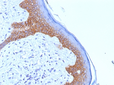
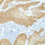
Variations in Desmoglein-3 antibody staining intensity in immunohistochemistry on tissue sections are present across different anatomical locations. Low, but measureable presence of Desmoglein-3 could be seen insquamous epithelial cells in the cervix, uterine, oral mucosa. We were unable to detect Desmoglein-3 in other tissues. Disease states, inflammation, and other physiological changes can have a substantial impact on antibody staining patterns. These measurements were all taken in tissues deemed normal or from patients without known disease.
| Desmoglein-3 General Information | |
|---|---|
| Alternate Names | |
| Desmoglein-3, DSG3 | |
| Molecular Weight | |
| 130kDa | |
| Chromosomal Location | |
| 18q12.1 | |
| Curated Database and Bioinformatic Data | |
| Gene Symbol | DSG3 |
| Entrez Gene ID | 1830 |
| Ensemble Gene ID | ENSG00000134757 |
| RefSeq Protein Accession(s) | XP_011524152, NP_001935 |
| RefSeq mRNA Accession(s) | XM_011525850, NM_001944 |
| RefSeq Genomic Accession(s) | NC_000018, NC_018929 |
| UniProt ID(s) | P32926 |
| UniGene ID(s) | P32926 |
| HGNC ID(s) | 3050 |
| Cosmic ID(s) | DSG3 |
| KEGG Gene ID(s) | hsa:1830 |
| PharmGKB ID(s) | PA27503 |
| General Description of Desmoglein-3. | |
| Recognizes a protein of 130kDa, identified as Desmoglein-3 (DSG3). This MAb is highly specific to Desmoglein-3 and does not cross-react with other members of the Desmoglein-family. DSG3 is a calcium-binding transmembrane glycoprotein component of desmosomes in vertebrate epithelial cells. Desmosomes are cell-cell junctions between epithelial, myocardial, and certain other cell types. Currently, three desmoglein subfamily members are identified and all are members of the cadherin cell adhesion molecule superfamily. | |



There are no reviews yet.