Human Anti-Filaggrin Antibody Product Attributes
Filaggrin Previously Observed Antibody Staining Patterns
Observed Subcellular, Organelle Specific Staining Data:
Anti-FLG antibody staining is expected to be primarily localized to the vesicles.
Observed Antibody Staining Data By Tissue Type:
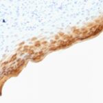
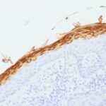
Variations in Filaggrin antibody staining intensity in immunohistochemistry on tissue sections are present across different anatomical locations. An intense signal was observed in epidermal cells in the skin. More moderate antibody staining intensity was present in epidermal cells in the skin. Low, but measureable presence of Filaggrin could be seen infibroblasts in skin, melanocytes in skin, squamous epithelial cells in the cervix and uterine. We were unable to detect Filaggrin in other tissues. Disease states, inflammation, and other physiological changes can have a substantial impact on antibody staining patterns. These measurements were all taken in tissues deemed normal or from patients without known disease.
| Filaggrin General Information | |
|---|---|
| Alternate Names | |
| FLG, Filaggrin, Filament Aggregating rotein | |
| Molecular Weight | |
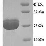
| 26-45kDa (Processed) ;350kDa (Profilaggrin) | |
| Chromosomal Location | |
| 1q21.3 | |
| Curated Database and Bioinformatic Data | |
| Gene Symbol | FLG |
| Entrez Gene ID | 2312 |
| Ensemble Gene ID | ENSG00000143631 |
| RefSeq Protein Accession(s) | NP_002007 |
| RefSeq mRNA Accession(s) | NM_002016, |
| RefSeq Genomic Accession(s) | NG_016190, NC_000001, NC_018912 |
| UniProt ID(s) | P20930 |
| UniGene ID(s) | P20930 |
| HGNC ID(s) | 3748 |
| Cosmic ID(s) | FLG |
| KEGG Gene ID(s) | hsa:2312 |
| PharmGKB ID(s) | PA28169 |
| General Description of Filaggrin. | |
| Filaggrin is an intermediate filament-associated protein that aggregates keratin intermediate filaments in mammalian epidermis. It is initially synthesized as a polyprotein precursor, profilaggrin (consisting of multiple filaggrin units of 324 aa each), which is localized in keratohyalin granules, and is subsequently proteolytically processed into individual functional filaggrin molecules. Active filaggrin is present at a level of the epidermis where keratinocytes are in transition between the live nucleated granular layer and the anucleate cornified layer, suggesting that filaggrin aids in the terminal differentiation process by facilitating apoptotic machinery. | |


There are no reviews yet.