

 PDF Datasheet
Human and Rat Anti-FOXA1 / HNF3A Antibody Product Attributes
FOXA1 / HNF3A Previously Observed Antibody Staining Patterns
Observed Subcellular, Organelle Specific Staining Data:
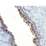
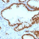
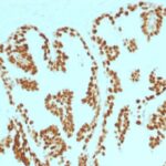
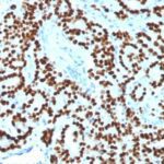
Anti-FOXA1 antibody staining is expected to be primarily localized to the nucleoli fibrillar center and nucleoplasm.
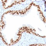
Observed Antibody Staining Data By Tissue Type:
Variations in FOXA1 / HNF3A antibody staining intensity in immunohistochemistry on tissue sections are present across different anatomical locations. An intense signal was observed in glandular cells in the prostate. More moderate antibody staining intensity was present in glandular cells in the prostate. Low, but measureable presence of FOXA1 / HNF3A could be seen inglandular cells in the appendix, breast, colon, duodenum, gallbladder, rectum and small intestine, squamous epithelial cells in the esophagus and oral mucosa and glandular cells in the appendix, breast, colon, duodenum, gallbladder and rectum and small intestine. We were unable to detect FOXA1 / HNF3A in other tissues. Disease states, inflammation, and other physiological changes can have a substantial impact on antibody staining patterns. These measurements were all taken in tissues deemed normal or from patients without known disease.
Observed Antibody Staining Data By Tissue Disease Status:
Tissues from cancer patients, for instance, have their own distinct pattern of FOXA1 / HNF3A expression as measured by anti-FOXA1 / HNF3A antibody immunohistochemical staining. The average level of expression by tumor is summarized in the table below. The variability row represents patient to patient variability in IHC staining.
| Sample Type | breast cancer | carcinoid | cervical cancer | colorectal cancer | endometrial cancer | glioma | head and neck cancer | liver cancer | lung cancer | lymphoma | melanoma | ovarian cancer | pancreatic cancer | prostate cancer | renal cancer | skin cancer | stomach cancer | testicular cancer | thyroid cancer | urothelial cancer |
|---|---|---|---|---|---|---|---|---|---|---|---|---|---|---|---|---|---|---|---|---|
| Signal Intensity | +++ | ++ | – | – | – | – | – | – | – | – | – | – | – | ++ | – | – | – | – | – | ++ |
| FOXA1 Variability | + | ++ | ++ | ++ | ++ | + | ++ | + | ++ | + | + | + | ++ | ++ | + | + | ++ | + | + | +++ |
| FOXA1 / HNF3A General Information | |
|---|---|
| Alternate Names | |
| Forkhead box protein A1, FOXA1, hepatocyte nuclear factor 3-alpha, HNF-3A, FOXA1 | |
| Molecular Weight | |
| 79kDa | |
| Chromosomal Location | |
| 14q21.1 | |
| Curated Database and Bioinformatic Data | |
| Gene Symbol | FOXA1 |
| Entrez Gene ID | 3169 |
| Ensemble Gene ID | ENSG00000129514 |
| RefSeq Protein Accession(s) | XP_016876735, NP_004487 |
| RefSeq mRNA Accession(s) | NM_004496 |
| RefSeq Genomic Accession(s) | NG_033028, NC_018925, NC_000014 |
| UniProt ID(s) | P55317 |
| UniGene ID(s) | P55317 |
| HGNC ID(s) | 5021 |
| Cosmic ID(s) | FOXA1 |
| KEGG Gene ID(s) | hsa:3169 |
| PharmGKB ID(s) | PA201090 |
| General Description of FOXA1 / HNF3A. | |
| The transcription factor Forkhead-box A1 (FOXA1), also known as hepatocyte nuclear factor 3-alpha, is a member of the FOX class of transcription factors. HNF-1 (? and ?), HNF-3 (?, ? and ?), HNF-4 (? and ?), and HNF-6 compose, in part, a homeoprotein family designated the hepatocyte nuclear factor family. The various HNF-1 isoforms regulate transcription of genes in the liver as well as in other tissues such as kidney, small intestine and thymus. FOXA1 is expressed in normal breast ductal epithelium and other epithelium in different organs, such as lung, pancreas, bladder, prostate, and colon. Recently, FOXA1 has been shown to be a major determinant of estrogen-ER activity and endocrine response in breast cancer cells. FOXA1 expression correlates with estrogen receptor (ER)-positivity, especially in luminal subtype A breast cancers, which is associated with favorable prognosis. FOXA1 is useful in the sub-classification of breast carcinomas. | |
There are no reviews yet.