


![Anti-/ Mouse Foxp3 Antibody [3G3] - Image 5](https://cdn-enquirebio.pressidium.com/wp-content/uploads/2017/10/enQuire-Bio-QAB71-PE-100ug-anti-FOXP3-antibody-10.png)

 PDF Datasheet
Mouse Anti-FOXP3 Antibody Product Attributes
FOXP3 Previously Observed Antibody Staining Patterns
Observed Subcellular, Organelle Specific Staining Data:
Staining with anti-FOXP3 antibody reveals FOXP3 expression is expected to be primarily localized to the nucleoplasm.
Observed Antibody Staining Data By Tissue Type:
Variations in FOXP3 / Scurfin antibody staining intensity in immunohistochemistry on tissue sections are present across different anatomical locations. Low, but measureable presence of FOXP3 / Scurfin could be seen inLeydig cells in the testis and macrophages in lung. We were unable to detect FOXP3 / Scurfin in other tissues. Disease states, inflammation, and other physiological changes can have a substantial impact on antibody staining patterns. These measurements were all taken in tissues deemed normal or from patients without known disease.
| FOXP3 General Information | |
|---|---|
| Alternate Names | |
| FOXP3, forkhead box P3, scurfin | |
| Curated Database and Bioinformatic Data | |
| Gene Symbol | Foxp3 |
| Entrez Gene ID | 50943, 20371 |
| Ensemble Gene ID | ENSG00000049768, ENSMUSG00000039521 |
| RefSeq Protein Accession(s) | XP_016885054, XP_016885056, XP_006724596, XP_016885055, NP_001107849, XP_011542218, NP_054728, NP_473380, NP_001186277, NP_001186276 |
| RefSeq mRNA Accession(s) | XM_011543916, XM_017029566, XM_006724533, XM_017029567, NM_001114377, NM_014009NM_054039, NM_001199347, NM_001199348 |
| RefSeq Genomic Accession(s) | NG_007392, NC_018934, NC_000023, NC_000086 |
| UniProt ID(s) | Q9BZS1, B7ZLG1, Q53Z59, Q99JB6 |
| UniGene ID(s) | Q9BZS1, B7ZLG1, Q53Z59, Q99JB6 |
| HGNC ID(s) | 6106 |
| Cosmic ID(s) | FOXP3, Foxp3 |
| KEGG Gene ID(s) | hsa:50943, mmu:20371 |
| PharmGKB ID(s) | PA201094 |
| General Description of FOXP3. | |
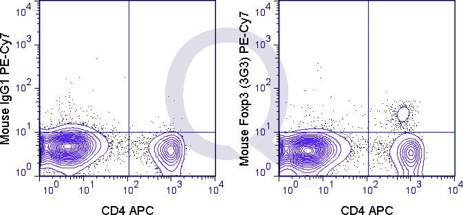
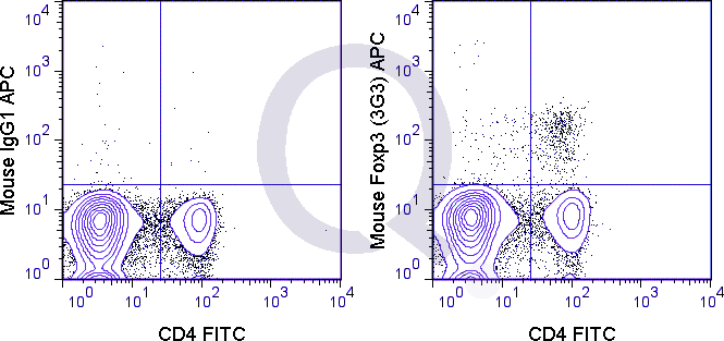
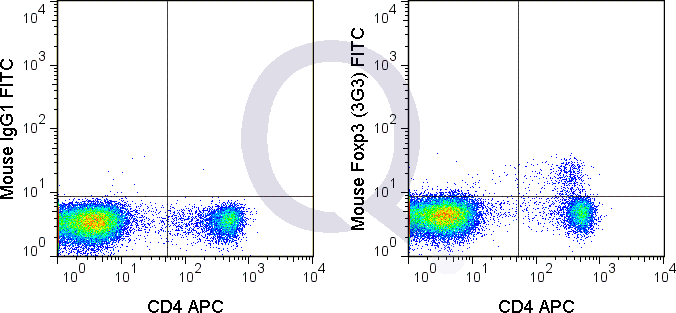
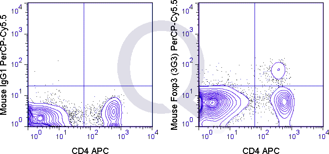
| The 3G3 antibody reacts with mouse Foxp3, a 50-55 kDa transcription factor which is a central regulator of T cell activity and is critical for the development and function of regulatory T cells (Tregs). Foxp3 is expressed at constitutively high levels in Treg cells, which are further identified as being CD4+ CD25+. In resting conventional T cells (CD4+ CD25-) Foxp3 expression is restricted, and upon TCR activation is expressed only transiently and in a small proportion of cells. However, the growth factor TGF-beta has been shown to induce expression of Foxp3 in nave T cells, driving their development into Foxp3+ Tregs, which are called induced or adaptive Tregs. These cells are phenotypically similar to so-called natural Tregs (CD4+ CD25high Foxp3+) which originate in the thymus and comprise the majority of Treg cells. Tregs are critical for maintaining peripheral tolerance and are implicated in the development of autoimmunity.It is important to review the literature in choosing an antibody for the Foxp3 antigen in flow cytometry, as the potential for high background or non-specific staining may be observed. The 3G3 antibody may be used for intracellular detection of Foxp3 in cells from mouse and Rhesus macaque. | |
Selected References
Limitations and Warranty
| Size | |
|---|---|
| Tag | APC, FITC, PE, PE-Cy7, PerCP-Cy5.5 |
| Buffer and Stabilizer | 10 mM NaH2PO4, 150 mM NaCl, 0.05% BSA, 0.05% NaN3, pH7.2, 10mM Tris, 150 mM NaCl, 0.05% BSA, 0.05% NaN3, pH8 |
| Product Type | |
| Host | |
| Isotype | |
| Applications | |
| Species | |
| Mass Spec Validated? |
Only logged in customers who have purchased this product may leave a review.
There are no reviews yet.