
.jpg)
 PDF Datasheet
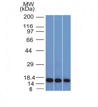
Human Anti-Galectin-1 Antibody Product Attributes
Galectin-1 Previously Observed Antibody Staining Patterns
Observed Subcellular, Organelle Specific Staining Data:
Anti-LGALS1 antibody staining is expected to be primarily localized to the cytosol and nucleoplasm. There is variability in either the signal strength or the localization of signal in cytosol, nucleoplasm and cytosol from cell to cell.
Observed Antibody Staining Data By Tissue Type:
Variations in Galectin-1 antibody staining intensity in immunohistochemistry on tissue sections are present across different anatomical locations. An intense signal was observed in adipocytes in mesenchymal tissue, cells in the endometrial stroma in endometrium, decidual cells in the placenta and smooth muscle cells in the smooth muscle. More moderate antibody staining intensity was present in adipocytes in mesenchymal tissue, cells in the endometrial stroma in endometrium, decidual cells in the placenta and smooth muscle cells in the smooth muscle. Low, but measureable presence of Galectin-1 could be seen in cells in the glomeruli in kidney, cells in the red pulp in spleen, cells in the seminiferous ducts in testis, cells in the white pulp in spleen, chondrocytes in mesenchymal tissue, fibroblasts in skin and mesenchymal tissue, germinal center cells in the lymph node, glandular cells in the parathyroid gland, glial cells in the caudate nucleus and hippocampus, hematopoietic cells in the bone marrow, keratinocytes in skin, Langerhans in skin, macrophages in lung, neuronal cells in the cerebral cortex and hippocampus, neuropil in cerebral cortex, non-germinal center cells in the lymph node and tonsil, pneumocytes in lung, squamous epithelial cells in the cervix and uterine and vagina. We were unable to detect Galectin-1 in other tissues. Disease states, inflammation, and other physiological changes can have a substantial impact on antibody staining patterns. These measurements were all taken in tissues deemed normal or from patients without known disease.
Observed Antibody Staining Data By Tissue Disease Status:
Tissues from cancer patients, for instance, have their own distinct pattern of Galectin-1 expression as measured by anti-Galectin-1 antibody immunohistochemical staining. The average level of expression by tumor is summarized in the table below. The variability row represents patient to patient variability in IHC staining.
| Sample Type | breast cancer | carcinoid | cervical cancer | colorectal cancer | endometrial cancer | glioma | head and neck cancer | liver cancer | lung cancer | lymphoma | melanoma | ovarian cancer | pancreatic cancer | prostate cancer | renal cancer | skin cancer | stomach cancer | testicular cancer | thyroid cancer | urothelial cancer |
|---|---|---|---|---|---|---|---|---|---|---|---|---|---|---|---|---|---|---|---|---|
| Signal Intensity | – | – | – | – | + | ++ | ++ | – | – | – | +++ | – | – | – | – | – | – | – | + | – |
| LGALS1 Variability | ++ | ++ | + | + | +++ | +++ | ++ | + | ++ | + | ++ | + | + | + | ++ | ++ | + | + | ++ | ++ |
| Galectin-1 General Information | |
|---|---|
| Alternate Names | |
| Galectin-1, LGALS1 | |
| Molecular Weight | |
| 14kDa | |
| Chromosomal Location | |
| 22q13.1 | |
| Curated Database and Bioinformatic Data | |
| Gene Symbol | LGALS1 |
| Entrez Gene ID | 3956 |
| Ensemble Gene ID | ENSG00000100097 |
| RefSeq Protein Accession(s) | NP_002296 |
| RefSeq mRNA Accession(s) | NM_002305 |
| RefSeq Genomic Accession(s) | NC_018933, NC_000022 |
| UniProt ID(s) | P09382 |
| UniGene ID(s) | P09382 |
| HGNC ID(s) | 6561 |
| Cosmic ID(s) | LGALS1 |
| KEGG Gene ID(s) | hsa:3956 |
| PharmGKB ID(s) | PA30337 |
| General Description of Galectin-1. | |
| Galectin-1 is a member of the beta-galactoside-binding family and is a dimeric protein of 14kD participating in a variety of normal and pathological processes, including cancer progression. Galectin-1 can affect the proliferation of normal and malignant cells. Inhibition of cell growth is observed in a lactose-dependent manner as lower concentrations of the lectin stimulate cell proliferation. Galectin-1 may also be implicated in the induction of apoptosis of activated T cells through the binding of exogenous galectin-1 to CD45 molecules present on the surface of lymphocytes. Galectin-1, reported to be present either at the surface of cancer cells or accumulated around these cells could act as an immunological shield to protect against a T cell immune response and provide an advantage for survival. | |
Reviews
There are no reviews yet.