
 PDF Datasheet
Human Anti-HLA-DRA Antibody Product Attributes
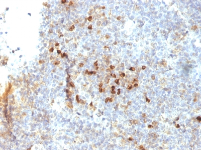
HLA-DRA Previously Observed Antibody Staining Patterns
Observed Antibody Staining Data By Tissue Type:
Variations in HLA-DRA antibody staining intensity in immunohistochemistry on tissue sections are present across different anatomical locations. An intense signal was observed in cells in the glomeruli in kidney, germinal center cells in the lymph node and tonsil, glandular cells in the duodenum and endometrium, Langerhans in skin, lymphoid tissue in appendix, macrophages in lung, myoepithelial cells in the breast and non-germinal center cells in the lymph node and tonsil. More moderate antibody staining intensity was present in cells in the glomeruli in kidney, germinal center cells in the lymph node and tonsil, glandular cells in the duodenum and endometrium, Langerhans in skin, lymphoid tissue in appendix, macrophages in lung, myoepithelial cells in the breast and non-germinal center cells in the lymph node and tonsil. Low, but measureable presence of HLA-DRA could be seen in cells in the red pulp in spleen, cells in the tubules in kidney, endothelial cells in the colon, epidermal cells in the skin, glandular cells in the appendix, cervix, uterine, epididymis and fallopian tube, glial cells in the caudate nucleus and hippocampus, myocytes in skeletal muscle, peripheral nerve in mesenchymal tissue, squamous epithelial cells in the tonsil and vagina and urothelial cells in the urinary bladder. We were unable to detect HLA-DRA in other tissues. Disease states, inflammation, and other physiological changes can have a substantial impact on antibody staining patterns. These measurements were all taken in tissues deemed normal or from patients without known disease.
| HLA-DRA General Information | |
|---|---|
| Alternate Names | |
| HLA class II histocompatibility antigen DR alpha chain, HLA-DRA | |
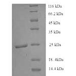
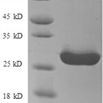
| Molecular Weight | |
| 36kDa (? chain), 27kDa (? chain) | |
| Chromosomal Location | |
| 6p21.3 | |
| Curated Database and Bioinformatic Data | |
| Gene Symbol | HLA-DRA |
| Entrez Gene ID | 3122 |
| Ensemble Gene ID | ENSG00000227993, ENSG00000204287, ENSG00000277263, ENSG00000230726, ENSG00000234794, ENSG00000228987, ENSG00000226260, ENSG00000206308 |
| RefSeq Protein Accession(s) | NP_061984 |
| RefSeq mRNA Accession(s) | NM_019111 |
| RefSeq Genomic Accession(s) | NC_000006, NG_187692, NG_167247, NG_113891, NC_018917, NG_167245, NG_167246, NG_167249, NG_002392, NG_167248 |
| UniProt ID(s) | P01903, A0A0G2JMH6 |
| UniGene ID(s) | P01903, A0A0G2JMH6 |
| HGNC ID(s) | 4947 |
| Cosmic ID(s) | HLA-DRA |
| KEGG Gene ID(s) | hsa:3122 |
| PharmGKB ID(s) | PA35071 |
| General Description of HLA-DRA. | |
| This MAb reacts with the HLA-DR antigen, a member of MHC class II molecules. It does not cross react with HLA-DP, HLA-DQ. HLA-DR is a heterodimeric cell surface glycoprotein comprised of a 36kDa alpha (heavy) chain, a 28kDa beta (light) chain. It is expressed on B-cells, activated T-cells, monocytes/macrophages, dendritic cells, other non-professional APCs. In conjunction with the CD3/TCR complex, CD4 molecules, HLA-DR is critical for efficient peptide presentation to CD4+ T cells. It is an excellent histiocytic marker in paraffin sections producing intense cytoplasmic staining. True histiocytic neoplasms are similarly positive. HLA-DR antigens also occur on a variety of epithelial cells, their corresponding neoplastic counterparts. | |
Reviews
There are no reviews yet.