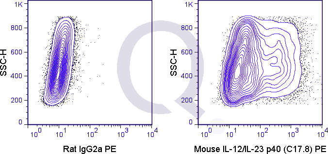
Mouse Anti-IL-12 p40 / IL-23 p40 Antibody Product Attributes
IL-12 p40 / IL-23 p40 Previously Observed Antibody Staining Patterns
Observed Antibody Staining Data By Tissue Type:
Variations in IL-12 p40 / IL-23 p40 antibody staining intensity in immunohistochemistry on tissue sections are present across different anatomical locations. Low, but measureable presence of IL-12 p40 / IL-23 p40 could be seen in cells in the seminiferous ducts in testis, cells in the tubules in kidney, glandular cells in the colon, duodenum, gallbladder, small intestine and stomach, glial cells in the caudate nucleus, hepatocytes in liver, keratinocytes in skin, myocytes in skeletal muscle and neuronal cells in the caudate nucleus. We were unable to detect IL-12 p40 / IL-23 p40 in other tissues. Disease states, inflammation, and other physiological changes can have a substantial impact on antibody staining patterns. These measurements were all taken in tissues deemed normal or from patients without known disease.
Observed Antibody Staining Data By Tissue Disease Status:
Tissues from cancer patients, for instance, have their own distinct pattern of IL-12 p40 / IL-23 p40 expression as measured by anti-IL-12 p40 / IL-23 p40 antibody immunohistochemical staining. The average level of expression by tumor is summarized in the table below. The variability row represents patient to patient variability in IHC staining.
| Sample Type | breast cancer | carcinoid | cervical cancer | colorectal cancer | endometrial cancer | glioma | head and neck cancer | liver cancer | lung cancer | lymphoma | melanoma | ovarian cancer | pancreatic cancer | prostate cancer | renal cancer | skin cancer | stomach cancer | testicular cancer | thyroid cancer | urothelial cancer |
|---|---|---|---|---|---|---|---|---|---|---|---|---|---|---|---|---|---|---|---|---|
| Signal Intensity | – | – | – | – | – | – | – | – | – | – | ++ | – | – | – | – | – | – | – | – | – |
| IL12B Variability | + | + | + | + | + | + | + | + | + | + | ++ | + | + | + | + | + | + | + | ++ | + |
| IL-12 p40 / IL-23 p40 General Information | |
|---|---|
| Alternate Names | |
| Interleukin-12, IL12 / Interleukin-23, IL23 p40, Cytotoxic lymphocyte maturation factor (CLMF), Natural killer cell stimulatory factor (NKSF), CTL maturation factor (TcMF), T-cell stimulating factor (TSF) | |
| Curated Database and Bioinformatic Data | |
| Gene Symbol | Il12b |
| Entrez Gene ID | 16160 |
| Ensemble Gene ID | ENSMUSG00000004296 |
| RefSeq Protein Accession(s) | NP_001290173 |
| RefSeq mRNA Accession(s) | NM_008352, NM_001303244 |
| RefSeq Genomic Accession(s) | NC_000077, |
| UniProt ID(s) | P43432, Q3ZAX5 |
| UniGene ID(s) | P43432, Q3ZAX5 |
| Cosmic ID(s) | Il12b |
| KEGG Gene ID(s) | mmu:16160 |
| General Description of IL-12 p40 / IL-23 p40. | |
| The C17.8 antibody is specific for the 40 kDa (p40) protein subunit shared by the cytokines IL-12 and IL-23. To form IL-12, p40 assembles with a separate 35 kDa protein known as p35, resulting in a 70 kDa functional cytokine. IL-12 is secreted by activated monocytes, macrophages, and dendritic cells, and has been shown to target nave, resting CD4+ T cells to promote their proliferation and secretion of cytokines. IL-23 contains the p40 subunit in combination with a 19 kDa protein chain, p19; its primary source being activated dendritic cells and other antigen-presenting cells. IL-23 appears to target different cell types than IL-12, acting on memory CD4+ T cells to induce a strong proliferative response and contributing to the generation and expansion of Th17 cells. Like the cytokines themselves, the receptors for IL-12 and IL-23 share one subunit, as well as containing distinct cytokine-specific subunits.As the C17.8 antibody binds to a shared subunit of both IL-12 and IL-23, it may be used as a marker for either IL-12 or IL-23 expression in dendritic cells, monocytes and macrophages, and is widely used for neutralization of activity associated with either cytokine. Please choose the appropriate format for each application. | |


There are no reviews yet.