
 PDF Datasheet
Antibody (Suitable for clinical applications)
| Specification | Recommendation |
|---|---|
| Recommended Dilution (Conc) | 1:50-1:100 |
| Pretreatment | EDTA Buffer pH8.0 |
| Incubation Parameters | 30 min at Room Temperature |
Prior to use, inspect vial for the presence of any precipitate or other unusual physical properties. These can indicate that the antibody has degraded and is no longer suitable for patient samples. Please run positive and negative controls simultaneously with all patient samples to account and control for errors in laboratory procedure. Use of methods or materials not recommended by enQuire Bio including change to dilution range and detection system should be routinely validated by the user.
Ki-67 Information for Pathologists
Summary:
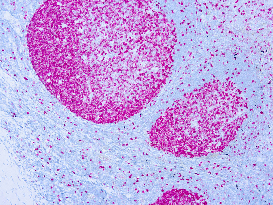
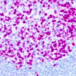
Marker of cell proliferation first described in 1983 (Int J Cancer 1983;31:13). Name derived from Kiel, Germany (city of origin) and the clone number in the 96 well plate (Wikipedia). Labile, nonhistone nuclear protein expressed in G1, S, G2 and M phases of cell cycle, then rapidly catabolized at end of M phase and not detectable in G0 and early G1 cells (J Cell Physiol 2000;182:311). MIB1 is the IgG1 antibody against Ki67 for formalin fixed, paraffin embedded tissue. Essential features
Common Uses By Pathologists:
Nuclear stain; cytoplasmic staining is disregarded. Distinguish benign / nonneoplastic and malignant / neoplastic lesions:. Anus / cervix: HSIL (Ki67+) versus atrophy or normal (Ki67-, Am J Surg Pathol 2010;34:1449). Colonic polyp with cautery artifact: distinguish adenoma (Ki67+) from nonadenomatous polyp (Ki67-, Arch Pathol Lab Med 2007;131:1089). Lymph node, sentinel node biopsy: distinguish melanoma (Ki67+) from nevus cells (Ki67-, Am J Surg Pathol 2002;26:1351).
| Ki-67 General Information | |
|---|---|
| Alternate Names | |
| Molecular Weight | |
| 358.7 kDa | |
| Chromosomal Location | |
| q26.2 [chr: 10] [chr_start: 128096659] [chr_end: 128126423] [strand: -1] | |
| Curated Database and Bioinformatic Data | |
| Gene Symbol | MKI67 |
| Entrez Gene ID | 4288 |
| RefSeq Protein Accession(s) | NP_001139438; NP_002408 |
| RefSeq mRNA Accession(s) | NM_002417; NM_001145966; XM_006717864; XM_011539818; |
| RefSeq Genomic Accession(s) | NG_047061; NC_000010 |
| UniProt ID(s) | P46013 |
| PharmGKB ID(s) | PA30825 |
| KEGG Gene ID(s) | hsa:4288 |
| General Description of Ki-67 . | |
| Ki-67 is a nuclear protein, which is expressed in proliferating cells. Ki-67 is preferentially expressed during late G1-, S-, M-, and G2-phases of the cell cycle, while cells in the G0 (quiescent) phase are negative for this protein. | |
Reviews
There are no reviews yet.