Human Anti-MAML3 Antibody Product Attributes
MAML3 Previously Observed Antibody Staining Patterns
Observed Subcellular, Organelle Specific Staining Data:
Anti-MAML3 antibody staining is expected to be primarily localized to the nuclear interchromatin granular clusters.
Observed Antibody Staining Data By Tissue Type:
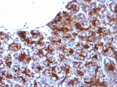
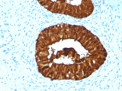
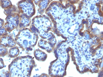
Variations in MAML3 antibody staining intensity in immunohistochemistry on tissue sections are present across different anatomical locations. An intense signal was observed in adipocytes in breast and mesenchymal tissue, chondrocytes in mesenchymal tissue, endothelial cells in the colon, epidermal cells in the skin, fibroblasts in skin and mesenchymal tissue, glandular cells in the colon, duodenum, gallbladder, rectum, salivary gland, stomach and thyroid gland, hematopoietic cells in the bone marrow, keratinocytes in skin, melanocytes in skin, peripheral nerve/ganglion in colon, squamous epithelial cells in the esophagus and oral mucosa and trophoblastic cells in the placenta. More moderate antibody staining intensity was present in adipocytes in breast and mesenchymal tissue, chondrocytes in mesenchymal tissue, endothelial cells in the colon, epidermal cells in the skin, fibroblasts in skin and mesenchymal tissue, glandular cells in the colon, duodenum, gallbladder, rectum, salivary gland, stomach and thyroid gland, hematopoietic cells in the bone marrow, keratinocytes in skin, melanocytes in skin, peripheral nerve/ganglion in colon, squamous epithelial cells in the esophagus and oral mucosa and trophoblastic cells in the placenta. Low, but measureable presence of MAML3 could be seen inbile duct cells in the liver, cells in the red pulp in spleen, germinal center cells in the tonsil, glandular cells in the parathyroid gland, glial cells in the caudate nucleus and hippocampus, neuronal cells in the caudate nucleus and cerebral cortex, peripheral nerve in mesenchymal tissue and Purkinje cells in the cerebellum. We were unable to detect MAML3 in other tissues. Disease states, inflammation, and other physiological changes can have a substantial impact on antibody staining patterns. These measurements were all taken in tissues deemed normal or from patients without known disease.
| MAML3 General Information | |
|---|---|
| Alternate Names | |
| MAML3, Mastermind Like Transcriptional Coactivator 3 | |
| Molecular Weight | |
| 150-170kDa | |
| Chromosomal Location | |
| 4q28 | |
| Curated Database and Bioinformatic Data | |
| Gene Symbol | MAML3 |
| Entrez Gene ID | 55534 |
| Ensemble Gene ID | ENSG00000196782 |
| RefSeq Protein Accession(s) | NP_061187 |
| RefSeq mRNA Accession(s) | NM_018717 |
| RefSeq Genomic Accession(s) | NC_000004, NC_018915 |
| UniProt ID(s) | Q9NPV6, Q96JK9 |
| UniGene ID(s) | Q9NPV6, Q96JK9 |
| HGNC ID(s) | 16272 |
| Cosmic ID(s) | MAML3 |
| KEGG Gene ID(s) | hsa:55534 |
| PharmGKB ID(s) | PA134953776 |
| General Description of MAML3. | |
| MAML3 (mastermind-like protein 3) is a nuclear speckle protein that acts as a transcriptional coactivator for Notch receptors. The Notch signaling pathway influences cell fate by regulating the ability of precursor cells to properly respond to developmental signals. MAML3 is a member of the mastermind-like family of proteins that are human homologs of the Drosophila melanogaster mastermind protein. Through its N-terminal region, MAML3 interacts with the ankyrin repeats of the Notch proteins Notch 1, Notch 2, Notch 3 and Notch 4.This interaction leads to formation of a DNA-binding complex with the Notch proteins and RBP-J? ;a complex that can then induce HES1 gene expression. While the N-terminal domain of MAML3 is essential for proper Notch binding, the C-terminal domain of MAML3 is essential for transcriptional activation. Due to its involvement in cell signaling and transcriptional activation, upregulation of MAML3 is thought to be involved in oncogenesis. | |


There are no reviews yet.