

![Analysis of Mass Spec data (dashed-line) of fractions stained with Moesin MS-QAVA™ monoclonal antibody [Clone: MSN/491] (solid-line), reveals that less than 0.8% of signal is attributable to non-specific binding of anti-Moesin [Clone: MSN/491] to targets other than MSN protein. Even frequently cited antibodies have much greater non-specific interactions, averaging over 30%. Data in image is from analysis in A431, RT4 and MCF7 cells.](https://cdn-enquirebio.pressidium.com/wp-content/uploads/2017/10/enQuire-Bio-4478-MSM1-P0-anti-Moesin-antibody.png)
 PDF Datasheet
Human and Rat Anti-Moesin Antibody Product Attributes
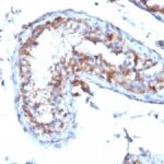
Moesin Previously Observed Antibody Staining Patterns
Observed Subcellular, Organelle Specific Staining Data:
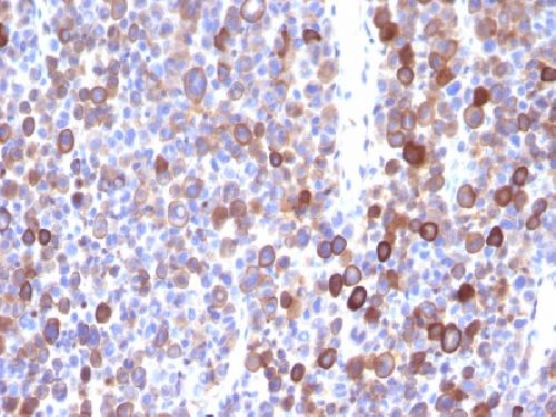
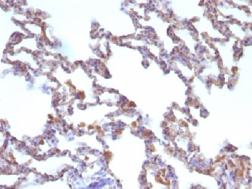
Variations in Moesin antibody staining intensity in immunohistochemistry on tissue sections are present across different anatomical locations. An intense signal was observed in hematopoietic cells in the bone marrow, germinal center cells in the lymph node and tonsil and non-germinal center cells in the tonsil. More moderate antibody staining intensity was present in hematopoietic cells in the bone marrow, germinal center cells in the lymph node and tonsil and non-germinal center cells in the tonsil. Low, but measureable presence of Moesin could be seen inglandular cells in the breast, cells in the molecular layer in cerebellum, glial cells in the cerebral cortex, glandular cells in the cervix, uterine, squamous epithelial cells in the cervix, uterine, cells in the endometrial stroma in endometrium, glandular cells in the endometrium and fallopian tube, follicle cells in the ovary, ovarian stroma cells in the ovary, exocrine glandular cells in the pancreas, glandular cells in the parathyroid gland, prostate and seminal vesicle, fibroblasts in skin, keratinocytes in skin, melanocytes in skin, smooth muscle cells in the smooth muscle, cells in the seminiferous ducts in testis, glandular cells in the thyroid gland and urothelial cells in the urinary bladder. We were unable to detect Moesin in other tissues. Disease states, inflammation, and other physiological changes can have a substantial impact on antibody staining patterns. These measurements were all taken in tissues deemed normal or from patients without known disease.
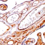
Observed Antibody Staining Data By Tissue Type:
Tissues from cancer patients, for instance, have their own distinct pattern of Moesin expression as measured by anti-Moesin antibody immunohistochemical staining. The average level of expression by tumor is summarized in the table below. The variability row represents patient to patient variability in IHC staining.
| Sample Type | breast cancer | carcinoid | cervical cancer | colorectal cancer | endometrial cancer | glioma | head and neck cancer | liver cancer | lung cancer | lymphoma | melanoma | ovarian cancer | pancreatic cancer | prostate cancer | renal cancer | skin cancer | stomach cancer | testicular cancer | thyroid cancer | urothelial cancer |
|---|---|---|---|---|---|---|---|---|---|---|---|---|---|---|---|---|---|---|---|---|
| Signal Intensity | – | – | ++ | – | + | – | +++ | + | ++ | +++ | ++ | + | ++ | – | ++ | ++ | – | + | +++ | – |
| MSN Variability | + | ++ | ++ | + | ++ | ++ | ++ | ++ | ++ | ++ | + | ++ | ++ | + | ++ | ++ | ++ | ++ | ++ | ++ |
| Moesin General Information | |
|---|---|
| Alternate Names | |
| HEL70, Moesin | |
| Molecular Weight | |
| 78kDa | |
| Chromosomal Location | |
| Xq11.1 | |
| Curated Database and Bioinformatic Data | |
| Gene Symbol | MSN |
| Entrez Gene ID | 4478 |
| Ensemble Gene ID | ENSG00000147065 |
| RefSeq Protein Accession(s) | XP_005262326, XP_011529261, XP_016885034, XP_016885035, NP_002435 |
| RefSeq mRNA Accession(s) | XM_005262269, XM_017029546, XM_017029545, XM_011530959 NM_002444 |
| RefSeq Genomic Accession(s) | NC_018934, NG_012516, NC_000023 |
| UniProt ID(s) | V9HWC0, P26038 |
| UniGene ID(s) | V9HWC0, P26038 |
| HGNC ID(s) | 7373 |
| Cosmic ID(s) | MSN |
| KEGG Gene ID(s) | hsa:4478 |
| PharmGKB ID(s) | PA31178 |
| General Description of Moesin. | |
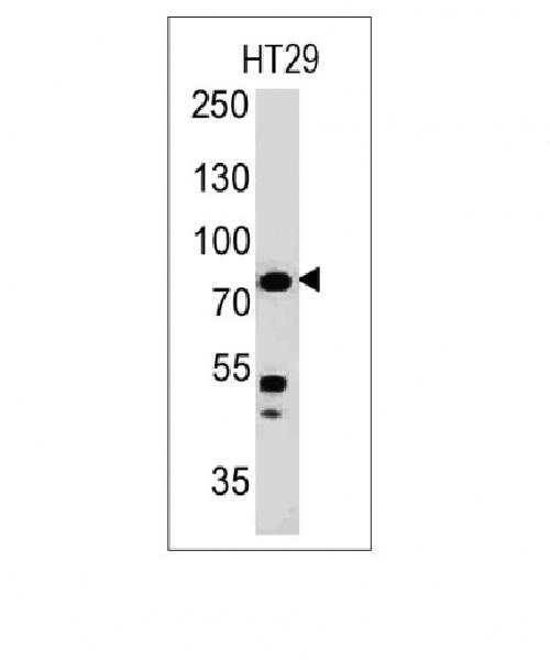
| Recognizes 78kDa moesin protein. Moesin, a member of the talin-4.1 superfamily, is a linking protein of the sub-membranous actin cytoskeleton. It is expressed in variable amounts in cells of different phenotypes such as macrophages, lymphocytes, fibroblastic, endothelial, epithelial, and neuronal cell lines but not in blood cells. The ERM proteins, ezrin, radixin, and moesin are involved in a variety of cellular functions, such as cell adhesion, migration, and the organization of cell surface structures, and are highly homologous, both in protein sequence and in functional activity, with merlin/schwannomin, a neurofibromatosis-2-associated tumor-suppressor protein. Cell lines of epithelial and mesothelial origin contain both moesin and radixin whereas cells of endothelial and lymphoid origin express moesin. | |
Reviews
There are no reviews yet.