
 PDF Datasheet
Human Anti-MUC1 / EMA / CD227 Antibody Product Attributes
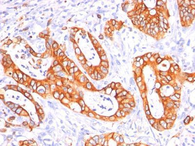
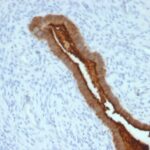
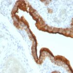
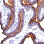
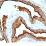
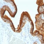
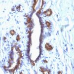
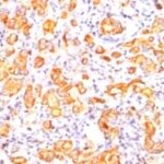
MUC1 / EMA / CD227 Previously Observed Antibody Staining Patterns
Observed Subcellular, Organelle Specific Staining Data:
Anti-MUC1 antibody staining is expected to be primarily localized to the plasma membrane and vesicles.
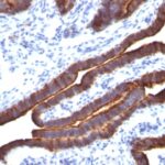
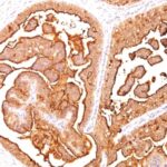
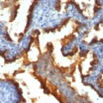
Observed Antibody Staining Data By Tissue Type:
Variations in MUC1 / EMA / CD227 antibody staining intensity in immunohistochemistry on tissue sections are present across different anatomical locations. An intense signal was observed in respiratory epithelial cells in the bronchus, glandular cells in the cervix, uterine, colon, endometrium, fallopian tube and gallbladder, respiratory epithelial cells in the nasopharynx and glandular cells in the rectum and stomach. More moderate antibody staining intensity was present in respiratory epithelial cells in the bronchus, glandular cells in the cervix, uterine, colon, endometrium, fallopian tube and gallbladder, respiratory epithelial cells in the nasopharynx and glandular cells in the rectum and stomach. Low, but measureable presence of MUC1 / EMA / CD227 could be seen inglandular cells in the adrenal gland, hematopoietic cells in the bone marrow, myoepithelial cells in the breast, squamous epithelial cells in the cervix, uterine, macrophages in lung, non-germinal center cells in the lymph node, squamous epithelial cells in the oral mucosa, glandular cells in the prostate and salivary gland, fibroblasts in skin, keratinocytes in skin, Langerhans in skin, melanocytes in skin, epidermal cells in the skin, cells in the seminiferous ducts in testis, Leydig cells in the testis and squamous epithelial cells in the tonsil. We were unable to detect MUC1 / EMA / CD227 in other tissues. Disease states, inflammation, and other physiological changes can have a substantial impact on antibody staining patterns. These measurements were all taken in tissues deemed normal or from patients without known disease.
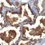
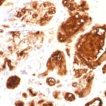
Observed Antibody Staining Data By Tissue Disease Status:
Tissues from cancer patients, for instance, have their own distinct pattern of MUC1 / EMA / CD227 expression as measured by anti-MUC1 / EMA / CD227 antibody immunohistochemical staining. The average level of expression by tumor is summarized in the table below. The variability row represents patient to patient variability in IHC staining.
| Sample Type | breast cancer | carcinoid | cervical cancer | colorectal cancer | endometrial cancer | glioma | head and neck cancer | liver cancer | lung cancer | lymphoma | melanoma | ovarian cancer | pancreatic cancer | prostate cancer | renal cancer | skin cancer | stomach cancer | testicular cancer | thyroid cancer | urothelial cancer |
|---|---|---|---|---|---|---|---|---|---|---|---|---|---|---|---|---|---|---|---|---|
| Signal Intensity | +++ | – | +++ | ++ | +++ | – | +++ | – | +++ | – | – | +++ | +++ | – | +++ | – | +++ | – | ++ | ++ |
| MUC1 Variability | + | ++ | + | ++ | + | + | ++ | ++ | ++ | + | + | + | ++ | + | ++ | ++ | + | ++ | ++ | ++ |
| MUC1 / EMA / CD227 General Information | |
|---|---|
| Alternate Names | |
| Mucin 1, cell surface associated, MUC1, polymorphic epithelial mucin, PEM | |
| Molecular Weight | |
| 265-400kDa | |
| Chromosomal Location | |
| 1q22 | |
| Curated Database and Bioinformatic Data | |
| Gene Symbol | MUC1 |
| Entrez Gene ID | 4582 |
| Ensemble Gene ID | ENSG00000185499 |
| RefSeq Protein Accession(s) | NP_001191214, NP_001191217, NP_001191219, NP_001191225, NP_001191216, NP_001191221, NP_001037857, NP_001037858, NP_001191224, NP_001191218, NP_001191222, NP_001191223, NP_002447, NP_001018016, NP_001018017, NP_001037855, NP_001037856, NP_001191215, NP_001191220, NP_001191226 |
| RefSeq mRNA Accession(s) | NM_001044392, NM_001204292, NM_002456, NM_001018021, NM_182741, NM_001204286, NM_001204293, NM_001204294, NM_001044390, NM_001044393, NM_001044391, NM_001204291, NM_001204295, NM_001018016, NM_001204285, NM_001204287, NM_001204290, NM_001204297 NM_001018017, NM_001204288, NM_001204289, NM_001204296 |
| RefSeq Genomic Accession(s) | NC_000001, NG_029383, NC_018912, |
| UniProt ID(s) | A0A0A0MRB3, A6ZIE6, A5YRU5, A6ZID7, A0A087X2A4, B6ECB3, A0A087X0L2, A0A0C4DGW3, Q7Z551, A6ZIE4, A6ZID6, A5YRV2, A5YRV0, Q7Z538, A5YRU7, P15941 |
| UniGene ID(s) | A0A0A0MRB3, A6ZIE6, A5YRU5, A6ZID7, A0A087X2A4, B6ECB3, A0A087X0L2, A0A0C4DGW3, Q7Z551, A6ZIE4, A6ZID6, A5YRV2, A5YRV0, Q7Z538, A5YRU7, P15941 |
| HGNC ID(s) | 7508 |
| Cosmic ID(s) | MUC1 |
| KEGG Gene ID(s) | hsa:4582 |
| PharmGKB ID(s) | PA31309 |
| General Description of MUC1 / EMA / CD227. | |
| In Western Blot, it recognizes proteins in MW range of 265-400kDa, identified as different glycoforms of EMA. The alpha subunit has cell adhesive properties. It can act both as an adhesion, an anti-adhesion protein. EMA may provide a protective layer on epithelial cells against bacterial, enzyme attack. The beta subunit contains a C-terminal domain, which is involved in cell signaling, through phosphorylations, protein-protein interactions. In immunohistochemical assays, it superbly stains routine formalin/paraffin carcinoma tissues. Antibody to EMA is useful as a pan-epithelial marker for detecting early metastatic loci of carcinoma in bone marrow or liver. | |
There are no reviews yet.