
 PDF Datasheet
Human Anti-MUC18 / CD146 / MCAM Antibody Product Attributes
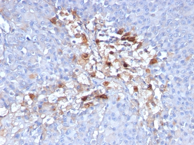
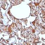
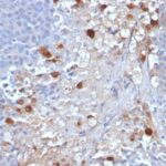
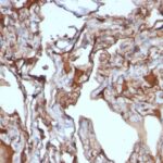
MUC18 / CD146 / MCAM Previously Observed Antibody Staining Patterns
Observed Subcellular, Organelle Specific Staining Data:
Anti-MCAM antibody staining is expected to be primarily localized to the plasma membrane.
Observed Antibody Staining Data By Tissue Type:
Variations in MUC18 / CD146 / MCAM antibody staining intensity in immunohistochemistry on tissue sections are present across different anatomical locations. An intense signal was observed in adipocytes in mesenchymal tissue, endothelial cells in the cerebral cortex and colon, fibroblasts in mesenchymal tissue, neuropil in cerebral cortex and peripheral nerve in mesenchymal tissue. More moderate antibody staining intensity was present in adipocytes in mesenchymal tissue, endothelial cells in the cerebral cortex and colon, fibroblasts in mesenchymal tissue, neuropil in cerebral cortex and peripheral nerve in mesenchymal tissue. Low, but measureable presence of MUC18 / CD146 / MCAM could be seen infibroblasts in skin, glial cells in the hippocampus, myocytes in heart muscle, neuronal cells in the cerebral cortex, Purkinje cells in the cerebellum and smooth muscle cells in the smooth muscle. We were unable to detect MUC18 / CD146 / MCAM in other tissues. Disease states, inflammation, and other physiological changes can have a substantial impact on antibody staining patterns. These measurements were all taken in tissues deemed normal or from patients without known disease.
Observed Antibody Staining Data By Tissue Disease Status:
Tissues from cancer patients, for instance, have their own distinct pattern of MUC18 / CD146 / MCAM expression as measured by anti-MUC18 / CD146 / MCAM antibody immunohistochemical staining. The average level of expression by tumor is summarized in the table below. The variability row represents patient to patient variability in IHC staining.
| Sample Type | breast cancer | carcinoid | cervical cancer | colorectal cancer | endometrial cancer | glioma | head and neck cancer | liver cancer | lung cancer | lymphoma | melanoma | ovarian cancer | pancreatic cancer | prostate cancer | renal cancer | skin cancer | stomach cancer | testicular cancer | thyroid cancer | urothelial cancer |
|---|---|---|---|---|---|---|---|---|---|---|---|---|---|---|---|---|---|---|---|---|
| Signal Intensity | – | – | – | – | + | – | + | – | + | – | ++ | – | – | – | – | – | – | + | – | – |
| MCAM Variability | ++ | ++ | ++ | + | ++ | ++ | ++ | ++ | ++ | + | ++ | + | ++ | + | + | ++ | + | ++ | ++ | + |
| MUC18 / CD146 / MCAM General Information | |
|---|---|
| Alternate Names | |
| CD146, cluster of differentiation 146, melanoma cell adhesion molecule, MCAM, cell surface glycoprotein MUC18 | |
| Molecular Weight | |
| 130kDa | |
| Chromosomal Location | |
| 11q23.3 | |
| Curated Database and Bioinformatic Data | |
| Gene Symbol | MCAM |
| Entrez Gene ID | 4162 |
| Ensemble Gene ID | ENSG00000076706 |
| RefSeq Protein Accession(s) | XP_016873248, NP_006491, XP_016873249, XP_016873250, XP_016873251 |
| RefSeq mRNA Accession(s) | XM_017017760, NM_006500, XM_017017759, XM_017017762, XM_017017761 |
| RefSeq Genomic Accession(s) | NC_018922, NC_000011 |
| UniProt ID(s) | P43121, A0A024R3I5 |
| UniGene ID(s) | P43121, A0A024R3I5 |
| HGNC ID(s) | 6934 |
| Cosmic ID(s) | MCAM |
| KEGG Gene ID(s) | hsa:4162 |
| PharmGKB ID(s) | PA30678 |
| General Description of MUC18 / CD146 / MCAM. | |
| The human CD146 gene maps to chromosome 11q23, encodes a trans-membrane glycoprotein, also designated MCAM, MUC 18, that belongs to the immunoglobulin superfamily, functions as a Ca2+-independent cell adhesion molecule. CD146 expression is restricted to advanced primary, metastatic melanomas, to cell lines of the neuroectodermal lineage, but not normal melanocytes. CD146 is found on 80% of advanced primary human melanomas, correlates well with development of metastatic disease. | |
Reviews
There are no reviews yet.